Mouse Siglec-E Antibody Summary
Gln20-Phe355
Accession # Q6PJ50
Applications
Please Note: Optimal dilutions should be determined by each laboratory for each application. General Protocols are available in the Technical Information section on our website.
Scientific Data
 View Larger
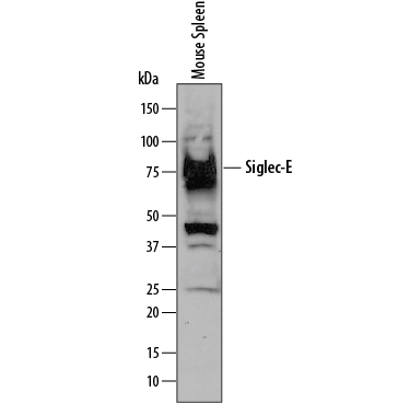
Detection of Mouse Siglec‑E by Western Blot. Western blot shows lysates of mouse spleen tissue. PVDF membrane was probed with 1 µg/mL of Goat Anti-Mouse Siglec-E Antigen Affinity-purified Polyclonal Antibody (Catalog # AF5806) followed by HRP-conjugated Anti-Goat IgG Secondary Antibody (Catalog # HAF109). A specific band was detected for Siglec-E at approximately 80 kDa (as indicated). This experiment was conducted under reducing conditions and using Immunoblot Buffer Group 1.
 View Larger
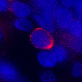
Siglec‑E in Mouse Spleen. Siglec-E was detected in perfusion fixed frozen sections of mouse spleen using Goat Anti-Mouse Siglec-E Antigen Affinity-purified Polyclonal Antibody (Catalog # AF5806) at 1.7 µg/mL overnight at 4 °C. Tissue was stained using the NorthernLights™ 557-conjugated Anti-Goat IgG Secondary Antibody (red; Catalog # NL001) and counterstained with DAPI (blue). Specific staining was localized to plasma membrane. View our protocol for Fluorescent IHC Staining of Frozen Tissue Sections.
Reconstitution Calculator
Preparation and Storage
- 12 months from date of receipt, -20 to -70 °C as supplied.
- 1 month, 2 to 8 °C under sterile conditions after reconstitution.
- 6 months, -20 to -70 °C under sterile conditions after reconstitution.
Background: Siglec-E
Siglecs are sialic acid specific I‑type lectins that are characterized by an extracellular domain (ECD) with an N‑terminal Ig‑like V‑set domain followed by varying numbers of Ig‑like C2‑set domains (1, 2). Mouse Siglec‑E, also known as Myeloid Inhibitory Siglec (MIS), is an 80 ‑ 85 kDa member of the CD33‑related subfamily of Siglecs. It consists of a 335 amino acid (aa) ECD with one Ig‑like V‑set domain and two Ig‑like C2‑set domains, a 21 aa transmembrane segment, and a 93 aa cytoplasmic domain that contains two immunoreceptor tyrosine‑based inhibitory motifs (ITIM) (3, 4). Rodent and primate Siglec gene families have significantly diverged, and Siglec‑9 is the most likely human ortholog of mouse Siglec‑E (1). Within the ECD, mouse Siglec‑E shares 56% and 80% aa sequence identity with human Siglec‑9 and rat Siglec‑E, respectively. Siglec‑E is expressed as a heavily N‑glycosylated disulfide‑linked homodimer and shows binding preference for disialic acids in the alpha 2‑8 linkage (3, 5). It is expressed on the surface of several hematopoietic cell types including neutrophils, NK cells, monocytes, peritoneal macrophages and B1 cells, and splenic myeloid dendritic cells and marginal zone B cells (5). Tyrosine phosphorylation of the cytoplasmic ITIMs mediates the association of Siglec‑E with the phosphatases SHP‑1 and SHP‑2 (3, 4). Siglec‑E is up‑regulated and additionally phosphorylated following cellular stimulation by a variety of TLR agonists (6). Siglec‑E signaling negatively regulates the LPS‑induced production of TNF‑ alpha and IL‑6 by macrophages (4, 6). Its up‑regulation in macrophages parallels the development of endotoxin tolerance (6). Siglec‑E recognition of sialylated determinants on virulent T. cruzi contributes to the suppression of dendritic cell IL‑12 p40 production (7).
- Varki, A. and T. Angata (2006) Glycobiology 16:1R.
- Crocker, P.R. et al. (2007) Nat. Rev. Immunol. 7:255.
- Yu, Z. et al. (2001) Biochem. J. 353:483.
- Ulyanova, T. et al. (2001) J. Biol. Chem. 276:14451.
- Zhang, J.Q. et al. (2004) Eur. J. Immunol. 34:1175.
- Boyd, C.R. et al. (2009) J. Immunol. 183:7703.
- Erdmann, H. et al. (2009) Cell. Microbiol. 11:1600.
Product Datasheets
Citations for Mouse Siglec-E Antibody
R&D Systems personnel manually curate a database that contains references using R&D Systems products. The data collected includes not only links to publications in PubMed, but also provides information about sample types, species, and experimental conditions.
5
Citations: Showing 1 - 5
Filter your results:
Filter by:
-
Siglec-7 restores ?-cell function and survival and reduces inflammation in pancreatic islets from patients with diabetes
Authors: G Dharmadhik, K Stolz, M Hauke, NG Morgan, A Varki, E de Koning, S Kelm, K Maedler
Sci Rep, 2017-04-05;7(0):45319.
Species: Human
Sample Types: Whole Cells
Applications: Functional Assay -
Studies on the detection, expression, glycosylation, dimerization and ligand binding properties of mouse Siglec-E
Authors: Shoib Siddiqui
J. Biol. Chem, 2016-12-05;0(0):.
Species: Hamster, Mouse
Sample Types: Whole Cells
-
Leishmania donovani Utilize Sialic Acids for Binding and Phagocytosis in the Macrophages through Selective Utilization of Siglecs and Impair the Innate Immune Arm
PLoS Negl Trop Dis, 2016-08-05;10(8):e0004904.
Species: Mouse
Sample Types: Cell Lysates
Applications: Western Blot -
Siglec receptors impact mammalian lifespan by modulating oxidative stress.
Authors: Schwarz F, Pearce O, Wang X, Samraj A, Laubli H, Garcia J, Lin H, Fu X, Garcia-Bingman A, Secrest P, Romanoski C, Heyser C, Glass C, Hazen S, Varki N, Varki A, Gagneux P
Elife, 2015-04-07;4(0):.
Species: Mouse
Sample Types: Whole Tissue
Applications: IHC-P -
Group B Streptococcus engages an inhibitory Siglec through sialic acid mimicry to blunt innate immune and inflammatory responses in vivo.
Authors: Chang Y, Olson J, Beasley F, Tung C, Zhang J, Crocker P, Varki A, Nizet V
PLoS Pathog, 2014-01-02;10(1):e1003846.
Species: Mouse
Sample Types: Cell Lysates
Applications: Western Blot
FAQs
No product specific FAQs exist for this product, however you may
View all Antibody FAQsReviews for Mouse Siglec-E Antibody
There are currently no reviews for this product. Be the first to review Mouse Siglec-E Antibody and earn rewards!
Have you used Mouse Siglec-E Antibody?
Submit a review and receive an Amazon gift card.
$25/€18/£15/$25CAN/¥75 Yuan/¥2500 Yen for a review with an image
$10/€7/£6/$10 CAD/¥70 Yuan/¥1110 Yen for a review without an image

