Mouse ST2/IL-33R APC-conjugated Antibody Summary
Ser27-Arg332
Accession # P14719
Applications
Please Note: Optimal dilutions should be determined by each laboratory for each application. General Protocols are available in the Technical Information section on our website.
Scientific Data
 View Larger
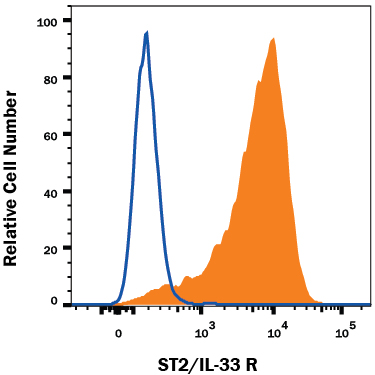
Detection of ST2/IL-33 R in P815 Mouse Cell Line by Flow Cytometry. P815 mouse mastocytoma cell line was stained with Rat Anti-Mouse ST2/IL-33 R APC-conjugated Monoclonal Antibody (Catalog # FAB10041A, filled histogram) or isotype control antibody (IC013A, open histogram). View our protocol for Staining Membrane-associated Proteins.
 View Larger
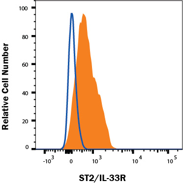
Detection of ST2/IL-33 R in Raw264.7 Mouse Cell Line by Flow Cytometry. Raw264.7 mouse monocyte/macrophage cell line was stained with Rat Anti-Mouse ST2/IL-33 R APC-conjugated Monoclonal Antibody (Catalog # FAB10041A, filled histogram) or isotype control antibody (IC013A, open histogram). Staining was performed using our Staining Membrane-associated Proteins protocol.
Reconstitution Calculator
Preparation and Storage
- 12 months from date of receipt, 2 to 8 °C as supplied.
Background: ST2/IL-33R
ST2, also known as IL-33R, IL-1 R4 and T1, is an Interleukin-1 receptor family glycoprotein that contributes to Th2 immune responses (1, 2). Mouse ST2 consists of a 306 amino acid (aa) extracellular domain (ECD) with three Ig-like domains, a 23 aa transmembrane segment, and a 212 aa cytoplasmic domain with an intracellular TIR domain (3). Alternate splicing of the 120 kDa mouse ST2 generates a soluble 60 kDa isoform that lacks the transmembrane and cytoplasmic regions (3). Within the ECD, mouse ST2 shares 68% and 81% aa sequence identity with human and rat ST2, respectively. ST2 is expressed on the surface of mast cells, activated Th2 cells, macrophages, and cardiac myocytes (4‑7). It binds IL-33, a cytokine that is up‑regulated by inflammation or mechanical strain in smooth muscle cells, airway epithelia, keratinocytes, and cardiac fibroblasts (4, 8). IL-33 binding induces the association of ST2 with IL-1R AcP, a shared signaling subunit that also associates with IL-1 RI and IL-1 R rp2 (1, 9, 10). In macrophages, ST2 interferes with signaling from IL-1 RI and TLR4 by sequestering the adaptor proteins MyD88 and Mal (6). In addition to its role in promoting mast cell and Th2 dependent inflammation, ST2 activation enhances antigen induced hypernociception and protects from atherosclerosis and cardiac hypertrophy (4, 11‑13). The soluble ST2 isoform is released by activated Th2 cells and strained cardiac myocytes and is elevated in the serum in allergic asthma (5, 7, 14). Soluble ST2 functions as a decoy receptor that blocks IL-33's ability to signal through transmembrane ST2 (9, 12‑14).
- Barksby, H.E. et al. (2007) Clin. Exp. Immunol. 149:217.
- Gadina, M. and C.A. Jefferies (2007) Science STKE 2007:pe31.
- Yanagisawa, K. et al. (1993) FEBS Lett. 318:83.
- Schmitz, J. et al. (2005) Immunity 23:479.
- Lecart, S. et al. (2002) Eur. J. Immunol. 32:2979.
- Brint, E.K. et al. (2004) Nat. Immunol. 5:373.
- Weinberg, E.O. et al. (2002) Circulation 106:2961.
- Sanada S. et al. (2007) J. Clin. Invest. 117:1538
- Palmer, G. et al. (2008) Cytokine 42:358.
- Chackerian, A.A. et al. (2007) J. Immunol. 179:2551.
- Allakhverdi, Z. et al. (2007) J. Immunol. 179:2051.
- Verri Jr., W.A. et al. (2008) Proc. Natl. Acad. Sci. 105:2723.
- Miller, A.M. et al. (2008) J. Exp. Med. 205:339.
- Hayakawa, H. et al. (2007) J. Biol. Chem. 282:26369.
Product Datasheets
Citations for Mouse ST2/IL-33R APC-conjugated Antibody
R&D Systems personnel manually curate a database that contains references using R&D Systems products. The data collected includes not only links to publications in PubMed, but also provides information about sample types, species, and experimental conditions.
3
Citations: Showing 1 - 3
Filter your results:
Filter by:
-
IL-33/ST2-mediated inflammation in macrophages is directly abrogated by IL-10 during rheumatoid arthritis
Authors: S Chen, B Chen, Z Wen, Z Huang, L Ye
Oncotarget, 2017-05-16;8(20):32407-32418.
Species: Mouse
Sample Types: Whole Cells
Applications: Flow Cytometry, ICC -
PD-1 regulates KLRG1(+) group 2 innate lymphoid cells
Authors: S Taylor, Y Huang, G Mallett, C Stathopoul, TC Felizardo, MA Sun, EL Martin, N Zhu, EL Woodward, MS Elias, J Scott, NJ Reynolds, WE Paul, DH Fowler, S Amarnath
J. Exp. Med., 2017-05-10;0(0):.
Species: Mouse
Sample Types: Whole Cells
Applications: Flow Cytometry -
Suppressive IL-17A(+)Foxp3(+) and ex-Th17 IL-17A(neg)Foxp3(+) Treg cells are a source of tumour-associated Treg cells
Authors: S Downs-Cann, S Berkey, GM Delgoffe, RP Edwards, T Curiel, K Odunsi, DL Bartlett, N Obermajer
Nat Commun, 2017-03-14;8(0):14649.
Species: Human
Sample Types: Whole Cells
Applications: Flow Cytometry
FAQs
No product specific FAQs exist for this product, however you may
View all Antibody FAQsReviews for Mouse ST2/IL-33R APC-conjugated Antibody
There are currently no reviews for this product. Be the first to review Mouse ST2/IL-33R APC-conjugated Antibody and earn rewards!
Have you used Mouse ST2/IL-33R APC-conjugated Antibody?
Submit a review and receive an Amazon gift card.
$25/€18/£15/$25CAN/¥75 Yuan/¥2500 Yen for a review with an image
$10/€7/£6/$10 CAD/¥70 Yuan/¥1110 Yen for a review without an image


