StemXVivo EMT Inducing Media Supplement (100X) Summary
To reliably induce epithelial to mesenchymal transition (EMT) in vitro.
Key Benefits
- Induces EMT in only 5 days
- Is compatible with multiple cell types
- Is a defined formulation to provide consistent EMT induction
Why Induce EMT in vitro with a Defined Media Supplement?
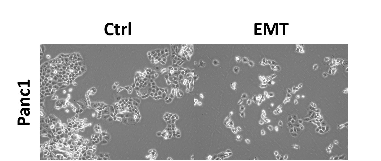
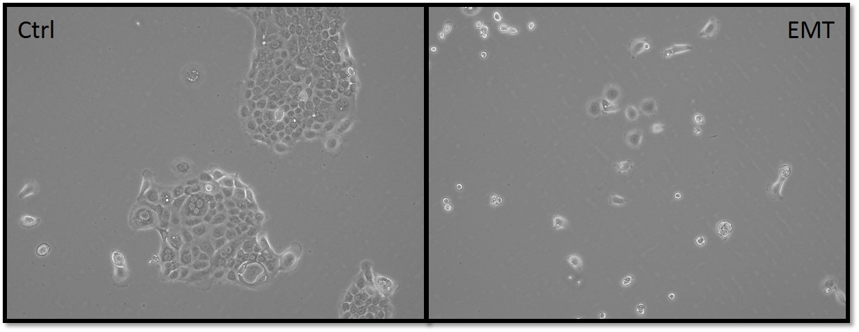
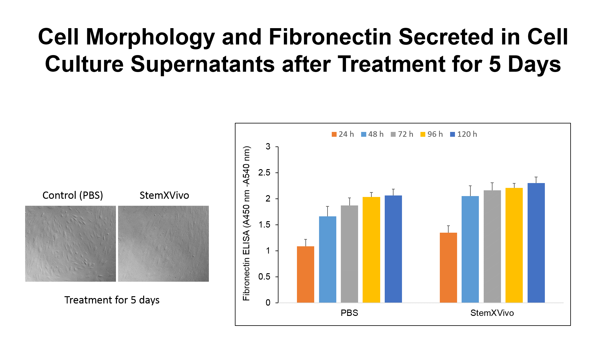
Designed to study EMT in vitro, this media supplement contains a cocktail of EMT-inducing recombinant proteins and neutralizing antibodies for the straightforward induction of EMT in a variety of cell types.
Reliable induction of EMT is useful for understanding the biology of EMT and for identifying and evaluating novel prognostic markers and therapeutics for fibrotic diseases and metastasis. However, the ability to induce EMT in vitro typically requires TGF-beta stimulation or labor-intensive genetic modification. Moreover, although TGF-beta is necessary to induce EMT, it is not sufficient in all cell types.
StemXVivo® EMT Inducing Media Supplement:
- Drives EMT in only 5 days to ensure efficient use of time and reagents.
- Provides a straightforward method to induce EMT in vitro.
- Is completely defined to reduce experimental variability.
- May induce EMT in cells resistant to TGF-beta stimulation alone.
- Avoids time-consuming genetic modifications.
Components
This defined media supplement contains the following premium quality proteins and high performance neutralizing antibodies to induce EMT:
- Recombinant Human Wnt-5a
- Recombinant Human TGF-beta1
- Anti-Human E-Cadherin
- Anti-Human sFRP-1
- Anti-Human Dkk-1
Each vial contains sufficient volume to drive EMT in the equivalent of fifteen 10 cm diameter tissue culture plates and has been shown to induce EMT in the following human cell lines:
- MCF-7 human breast cancer cells
- MCF-10A human breast epithelial cells
- HT-29 human colon adenocarcinoma cells
- A549 human lung carcinoma cells
- A431 human epithelial carcinoma cells
Specifications
Product Datasheets
Scientific Data
 View Larger
Induction of Epithelial to Mesenchymal Transition(EMT) in the A549 human lung carcinoma cell line. The A549 human lung carcinoma cell line was cultured without(A, B); or with(C, D)the StemXVivo®EMT Inducing Media Supplement (Catalog #CCM017) for five days.(A)Control cells exhibit typical epithelial morphology compared to the spindle-shaped, mesenchymal morphology observed in cells following EMT induction(C). EMT status was also assessed by dual immunofluorescence.(B)Untreated cells express the epithelial marker E-Cadherin (red) and exhibit little labeling of the mesenchymal marker Fibronectin (green).D)In contrast, EMT induction results in decreased E-Cadherin expression (red) and an increase in Fibronectin labeling (green). E-Cadherin was detected in cells using a NorthernLights™ (NL) 577-conjugated Goat Anti-Human E-Cadherin Antigen Affinity-purified Polyclonal Antibody (Catalog # NL648R; red). Fibronectin was detected using a Mouse Anti-Human Fibronectin Monoclonal Antibody (Catalog # MAB1918), followed by a NL493-conjugated Donkey Anti-Mouse IgG Secondary Antibody (Catalog # NL009; green). The nuclei were counterstained with DAPI (blue).
 View Larger
Upregulation of the Mesenchymal Markers, Snail and Vimentin, in EMT-induced Cells. A549 human lung carcinoma and MCF 10A human breast epithelial cells were either untreated or cultured with the StemXVivo®EMT Inducing Media Supplement (Catalog # CCM017) for five days. The cells were analyzed for an epithelial to mesenchymal transition (EMT) by simultaneously staining with the antibodies contained in the Human EMT 3-Color Immunocytochemistry Kit (Catalog # SC026): NorthernLights™ (NL) 637-conjugated Goat Anti-Human E-Cadherin (pseudocolored white), NL557-conjugated Goat Anti-Human Snail (red), and NL493-conjugated Rat Anti-Human Vimentin (green). Cells cultured with the EMT Inducing Media Supplement showed downregulation of the epithelial marker, E-Cadherin, and concurrent upregulation of the mesenchymal markers, Snail and Vimentin, compared to control cells.
Assay Procedure
Refer to the product datasheet for complete product details.
Briefly, an epithelial to mesenchymal transition (EMT) is induced using the following in vitro differentiation procedure:
- Add EMT Inducing Media Supplement directly to the standard culture media of your cells of interest
- Culture cells for only 5 days to induce EMT
- Evaluate EMT status using characteristic morphological changes and marker antibodies
Reagents supplied in the StemXVivo® EMT Inducing Media Supplement (Catalog # CCM017):
- 1 mL of StemXVivo® EMT Inducing Media Supplement
Note: The quantity of media supplement is sufficient to drive EMT in the equivalent of fifteen 10 cm diameter tissue culture plates.
Reagents
- Cell culture medium
- Dissociation solution (e.g., TrypLE™ Express, Invitrogen® or equivalent)
- 0.4% Trypan blue solution
Materials
- Epithelial cells of interest
- Tissue culture plates/flasks
- 15 mL centrifuge tubes
- Pipettes and pipette tips
- Serological pipettes
Equipment
- 37 °C and 5% CO2 incubator
- Centrifuge
- Hemocytometer
- Inverted microscope
- 37 °C water bath
- 2 °C to 8 °C refrigerator
Harvest epithelial cells of interest using a dissociation solution.

Transfer the cell suspension to a 15 mL conical tube containing pre-warmed media.
Centrifuge at 400 x g for 5 minutes.

Perform a cell count.

Plate cells at 0.9 - 1.0 x 10⁴ cells/cm2 in media containing 1X EMT Inducing Media Supplement.

Culture cells and monitor cell morphology daily.
Replace media after 3 days.

After 5 days, EMT induction is complete.
Evaluate EMT status using established markers and immunocytochemistry

Citations for StemXVivo EMT Inducing Media Supplement (100X)
R&D Systems personnel manually curate a database that contains references using R&D Systems products. The data collected includes not only links to publications in PubMed, but also provides information about sample types, species, and experimental conditions.
8
Citations: Showing 1 - 8
Filter your results:
Filter by:
-
Epithelial plasticity in COPD results in cellular unjamming due to an increase in polymerized actin
Authors: B Ghosh, K Nishida, L Chandrala, S Mahmud, S Thapa, C Swaby, S Chen, AA Khosla, J Katz, VK Sidhaye
Journal of Cell Science, 2022-02-24;0(0):. 2022-02-24
-
Immune-Stimulatory Effects of Curcumin on the Tumor Microenvironment in Head and Neck Squamous Cell Carcinoma
Authors: C Kötting, L Hofmann, R Lotfi, D Engelhardt, S Laban, PJ Schuler, TK Hoffmann, C Brunner, MN Theodoraki
Cancers, 2021-03-16;13(6):. 2021-03-16
-
Long non-coding RNA LCAL62 / LINC00261 is associated with lung adenocarcinoma prognosis
Authors: HX Dang, NM White, EB Rozycki, BM Felsheim, MA Watson, R Govindan, J Luo, CA Maher
Heliyon, 2020-03-05;6(3):e03521. 2020-03-05
-
Metastatic renal cell carcinoma cells growing in 3D on poly?D?lysine or laminin present a stem?like phenotype and drug resistance
Authors: KK Brodaczews, ZF Bielecka, K Maliszewsk, C Szczylik, C Porta, E Bartnik, AM Czarnecka
Oncol. Rep., 2019-09-18;42(5):1878-1892. 2019-09-18
-
Assessing metastatic potential of breast cancer cells based on EGFR dynamics
Authors: YL Liu, CK Chou, M Kim, R Vasisht, YA Kuo, P Ang, C Liu, EP Perillo, YA Chen, K Blocher, H Horng, YI Chen, DT Nguyen, TE Yankeelov, MC Hung, AK Dunn, HC Yeh
Sci Rep, 2019-03-04;9(1):3395. 2019-03-04
-
E-Cadherin in Colorectal Cancer: Relation to Chemosensitivity
Authors: I Druzhkova, N Ignatova, N Prodanets, N Kiselev, I Zhukov, M Shirmanova, V Zagainov, E Zagaynova
Clin Colorectal Cancer, 2018-10-16;0(0):. 2018-10-16
-
Thymidylate synthase is functionally associated with ZEB1 and contributes to the epithelial-to-mesenchymal transition of cancer cells
Authors: A Siddiqui, ME Vazakidou, A Schwab, F Napoli, C Fernandez-, I Rapa, MP Stemmler, M Volante, T Brabletz, P Ceppi
J. Pathol, 2017-06-01;0(0):. 2017-06-01
-
Paracrine and autocrine signals induce and maintain mesenchymal and stem cell states in the breast.
Authors: Scheel C, Eaton E, Li S, Chaffer C, Reinhardt F, Kah K, Bell G, Guo W, Rubin J, Richardson A, Weinberg R
Cell, 2011-06-10;145(6):926-40. 2011-06-10
FAQs
-
What is the composition of the medium used to culture MCF-7 cells for the data shown on the StemXVivo Media supplement (100X), Catalog # CCM017 product insert?
The medium used to cuture MCF-7 cells for the data shown on Catalog # CCM017 is MEM containing NEAA, Sodium Pyruvate, 10% FBS, 2mM L-Glutamine, and Penicillin-Streptomycin
-
Has StemXVivo Media supplement (100X), Catalog # CCM017, been tested on mouse epithelial cells?
Elaborate testing of Catlaog # CCM017 with mouse epithelial cells has not been performed. The supplement was tested once with NMuMG mouse mammary epithelial cells and found to induce EMT.
Reviews for StemXVivo EMT Inducing Media Supplement (100X)
Average Rating: 4.5 (Based on 6 Reviews)
Have you used StemXVivo EMT Inducing Media Supplement (100X)?
Submit a review and receive an Amazon gift card.
$25/€18/£15/$25CAN/¥75 Yuan/¥2500 Yen for a review with an image
$10/€7/£6/$10 CAD/¥70 Yuan/¥1110 Yen for a review without an image
Filter by:
It seems very useful, but too expensive. I gave up using it because of the high cost.


