
We offer a 100% guarantee on all our products.
Learn MoreILC2 Cell Markers
Click on one of the ILC subsets shown in the buttons below to see the markers that are commonly used to identify each cell type.

CD1a-
CD3-
CD11c-
CD14-
CD19-
CD34-
CD94-
DLEC/CLEC4C/BDCA-2-
Fc gamma RIII/CD16-
Fc epsilon RI-
IL-3 R alpha/CD123-
TCR alpha/beta-
TCR gamma/delta-
MOUSE
CD3-
B220/CD45 R-
CD5-
CD11b-
CD19-
Gr-1/Ly6G-
Ter-119-
A note regarding Lin- Markers
Overview
Group 2 ILCs (ILC2s) are a subset of innate lymphoid cells (ILCs) that has been suggested to be the innate counterpart of Th2 cells. Similar to Th2 cells, ILC2s are involved in the immune response against large extracellular pathogens such as helminths, and they promote type 2 inflammation and tissue repair. In addition, they require the expression of the transcription factor GATA-3 for their maintenance and survival, and they produce Amphiregulin, IL-4, IL-5, IL-9, and IL-13 following infection. ILC2s are activated by IL-25, IL-33, IL-4, TSLP, and prostaglandin D2 (PGD2), and like ILC1s and ILC3s, they are primarily localized to mucosal tissues and lack both lineage markers that define other immune cell types and rearranged antigen receptors. Exaggerated ILC2 immune responses are associated with atopic diseases such as asthma, chronic rhinosinusitis, and atopic dermatitis. In mice, ILC2s have been described as Lin-Sca-1+CD127+CD117+ST2+ICOS+ cells that also typically express CD90/Thy1, IL-17 RB, and CD25/IL-2 R alpha. In humans, ILC2s are commonly identified as Lin-CD127+CD45highCD161+CRTH-2+ cells that also express ST2 and IL-17 RB.
Data Examples
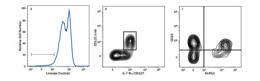
Detection of Cell Surface Markers on ILCs from Mouse Peyer’s Patches by Flow Cytometry. (A) Single cell suspensions were obtained from mouse Peyer’s patches and stained with a mouse lineage marker cocktail containing Alexa Fluor®700-conjugated monoclonal antibodies against CD3, B220, Ter-119, CD11b/Integrin alpha M, Gr-1, and CD5 (R&D Systems, Catalog # FLC001N). Lineage negative cells (CD3-, B220-, Ter-119-, CD11b/Integrin alpha M-, Gr-1-, and CD5-) were gated on by flow cytometry. (B) CD117+ CD127+ ILCs in the lineage negative population were detected by staining with an APC-conjugated Rat Anti-Mouse CD117/c-kit Monoclonal Antibody (R&D Systems, Catalog # FAB1356A) and an Alexa Fluor®488-conjugated Rat Anti-Mouse CD127/IL-7 R alpha Monoclonal Antibody (R&D Systems, Catalog # FAB47742G). (C) KLRG1 and CD25/IL-2 R alpha were detected on Lin- CD117+ CD127+ ILCs by staining with a PE-conjugated Rabbit Anti-Mouse KLRG1 Monoclonal Antibody (R&D Systems, Catalog # FAB6944P) and a PerCP-conjugated Rat Anti-Mouse CD25/IL-2 R alpha Monoclonal Antibody (R&D Systems, Catalog # FAB2438C).