Human EGFR Biotinylated Antibody Summary
Leu25-Ser645
Accession # CAA25240
Applications
Human EGFR Sandwich Immunoassay
Please Note: Optimal dilutions should be determined by each laboratory for each application. General Protocols are available in the Technical Information section on our website.
Scientific Data
 View Larger
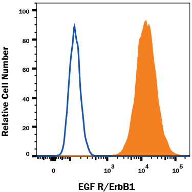
Detection of EGFR in A431 Human Cell Line by Flow Cytometry. A431 human epithelial carcinoma cell line was stained with Human EGFR Biotinylated Antigen Affinity-purified Polyclonal Antibody (Catalog # BAF231, filled histogram) or control antibody (Catalog # BAF108, open histogram), followed by Streptavidin-Allophycocyanin (Catalog # F0050).
Reconstitution Calculator
Preparation and Storage
- 12 months from date of receipt, -20 to -70 °C as supplied.
- 1 month, 2 to 8 °C under sterile conditions after reconstitution.
- 6 months, -20 to -70 °C under sterile conditions after reconstitution.
Background: EGFR
The epidermal growth factor receptor (EGFR) subfamily of receptor tyrosine kinases comprises four members: EGFR (also known as HER1, ErbB1 or ErbB), ErbB2 (Neu, HER2), ErbB3 (HER3), and ErbB4 (HER4). All family members are type I transmembrane glycoproteins that have an extracellular domain which contains two cysteine-rich domains separated by a spacer region that is involved in ligand binding, and a cytoplasmic domain which has a membrane-proximal tyrosine kinase domain and a C-terminal tail with multiple tyrosine autophosphorylation sites. The human EGFR gene encodes a 1210 amino acid (aa) residue precursor with a 24 aa putative signal peptide, a 621 aa extracellular domain, a 23 aa transmembrane domain, and a 542 aa cytoplasmic domain. EGFR has been shown to bind a subset of the EGF family ligands, including EGF, amphiregulin, TGF-alpha, betacellulin, epiregulin, heparin-binding EGF and neuregulin-2 alpha in the absence of a co-receptor. Ligand binding induces EGFR homodimerization as well as heterodimerization with ErbB2, resulting in kinase activation, tyrosine phosphorylation and cell signaling. EGFR can also be recruited to form heterodimers with the ligand-activated ErbB3 or ErbB4. EGFR signaling has been shown to regulate multiple biological functions including cell proliferation, differentiation, motility and apoptosis. In addition, EGFR signaling has also been shown to play a role in carcinogenesis (1-3).
- Daly, R.J. (1999) Growth Factors, 16:255.
- Schlessinger, J. (2000) Cell. 103:211.
- Maihle, N.J. et al. (2002) Cancer Treat. Res. 107:247.
Product Datasheets
Citations for Human EGFR Biotinylated Antibody
R&D Systems personnel manually curate a database that contains references using R&D Systems products. The data collected includes not only links to publications in PubMed, but also provides information about sample types, species, and experimental conditions.
5
Citations: Showing 1 - 5
Filter your results:
Filter by:
-
Magnetic Nanowire Networks for Dual-Isolation and Detection of Tumor-Associated Circulating Biomarkers
Authors: H Lee, M Choi, J Lim, M Jo, JY Han, TM Kim, Y Cho
Theranostics, 2018-01-01;8(2):505-517.
Applications: Functional Assay -
Activation of EGFR by small compounds through coupling the generation of hydrogen peroxide to stable dimerization of Cu/Zn SOD1.
Authors: Sakanyan V, Hulin P, Alves de Sousa R, Silva V, Hambardzumyan A, Nedellec S, Tomasoni C, Loge C, Pineau C, Roussakis C, Fleury F, Artaud I
Sci Rep, 2016-02-17;6(0):21088.
Species: Human
Sample Types: Cell Lysates
Applications: Western Blot -
SH2-PLA: a sensitive in-solution approach for quantification of modular domain binding by proximity ligation and real-time PCR.
Authors: Thompson C, Bloom L, Ogiue-Ikeda M, Machida K
BMC Biotechnol, 2015-06-26;15(0):60.
Species: Human
Sample Types: Cell Lysates
Applications: Functional Assay -
Protein shedding in urothelial bladder cancer: prognostic implications of soluble urinary EGFR and EpCAM.
Authors: Bryan R, Regan H, Pirrie S, Devall A, Cheng K, Zeegers M, James N, Knowles M, Ward D
Br J Cancer, 2015-03-17;112(6):1052-8.
Species: Human
Sample Types: Cell Lysates
Applications: Western Blot -
Development and validation of sandwich ELISA microarrays with minimal assay interference.
Authors: Gonzalez RM, Seurynck-Servoss SL, Crowley SA
J. Proteome Res., 2008-04-19;7(6):2406-14.
Species: Human
Sample Types: Serum
Applications: ELISA Microarray Development
FAQs
No product specific FAQs exist for this product, however you may
View all Antibody FAQsReviews for Human EGFR Biotinylated Antibody
Average Rating: 4.3 (Based on 3 Reviews)
Have you used Human EGFR Biotinylated Antibody?
Submit a review and receive an Amazon gift card.
$25/€18/£15/$25CAN/¥75 Yuan/¥2500 Yen for a review with an image
$10/€7/£6/$10 CAD/¥70 Yuan/¥1110 Yen for a review without an image
Filter by:


