Human/Primate EGF Antibody Summary
Applications
Human/Primate EGF Sandwich Immunoassay
Please Note: Optimal dilutions should be determined by each laboratory for each application. General Protocols are available in the Technical Information section on our website.
Scientific Data
 View Larger
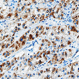
EGF in Human Lung Cancer Tissue. EGF was detected in immersion fixed paraffin-embedded sections of human lung cancer tissue using Mouse Anti-Human/Primate EGF Monoclonal Antibody (Catalog # MAB636) at 15 µg/mL overnight at 4 °C. Tissue was stained using the Anti-Mouse HRP-DAB Cell & Tissue Staining Kit (brown; Catalog # CTS002) and counterstained with hematoxylin (blue). Specific staining was localized to the plasma membrane and cytoplasm. View our protocol for Chromogenic IHC Staining of Paraffin-embedded Tissue Sections.
Reconstitution Calculator
Preparation and Storage
- 12 months from date of receipt, -20 to -70 °C as supplied.
- 1 month, 2 to 8 °C under sterile conditions after reconstitution.
- 6 months, -20 to -70 °C under sterile conditions after reconstitution.
Background: EGF
EGF is the prototypic member of a family of growth factors that are characterized by the presence of EGF like domains and activate members of the EGF receptor family. Proteolytic cleavage of a membrane-bound precursor releases mature soluble EGF which interacts with the EGF R to promote proliferation and differentiation of mesenchymal and epithelial cells.
Product Datasheets
Citations for Human/Primate EGF Antibody
R&D Systems personnel manually curate a database that contains references using R&D Systems products. The data collected includes not only links to publications in PubMed, but also provides information about sample types, species, and experimental conditions.
2
Citations: Showing 1 - 2
Filter your results:
Filter by:
-
Pre-analytical effects of blood sampling and handling in quantitative immunoassays for rheumatoid arthritis.
Authors: Zhao X, Qureshi F, Eastman PS, Manning WC, Alexander C, Robinson WH, Hesterberg LK
J. Immunol. Methods, 2012-02-17;378(1):72-80.
Species: Human
Sample Types: Serum
Applications: ELISA Development -
Accumulation of hEGF and hEGF-fusion proteins in chloroplast-transformed tobacco plants is higher in the dark than in the light.
Authors: Wirth S, Segretin ME, Mentaberry A, Bravo-Almonacid F
J. Biotechnol., 2006-04-03;125(2):159-72.
Species: Human
Sample Types: Cell Lysates, Tissue Homogenates
Applications: ELISA Development, Western Blot
FAQs
No product specific FAQs exist for this product, however you may
View all Antibody FAQsReviews for Human/Primate EGF Antibody
Average Rating: 5 (Based on 2 Reviews)
Have you used Human/Primate EGF Antibody?
Submit a review and receive an Amazon gift card.
$25/€18/£15/$25CAN/¥75 Yuan/¥2500 Yen for a review with an image
$10/€7/£6/$10 CAD/¥70 Yuan/¥1110 Yen for a review without an image
Filter by:


