Human EGFR Antibody Summary
Leu25-Ser645
Accession # CAA25240
Applications
Human EGFR Sandwich Immunoassay
Please Note: Optimal dilutions should be determined by each laboratory for each application. General Protocols are available in the Technical Information section on our website.
Scientific Data
 View Larger
Detection of Human EGFR by Western Blot. Western blot shows lysates of HeLa human cervical epithelial carcinoma cell line and MDA-MB-231 human breast cancer cell line. PVDF membrane was probed with 1 µg/mL of Goat Anti-Human EGFR Antigen Affinity-purified Polyclonal Antibody (Catalog # AF231) followed by HRP-conjugated Anti-Goat IgG Secondary Antibody (Catalog # HAF017). A specific band was detected for EGFR at approximately 175 kDa (as indicated). This experiment was conducted under reducing conditions and using Immunoblot Buffer Group 1.
 View Larger
Detection of EGFR in A431 Human Cell Line by Flow Cytometry. A431 human epithelial carcinoma cell line was stained with Goat Anti-Human EGFR Antigen Affinity-purified Polyclonal Antibody (Catalog # AF231, filled histogram) or isotype control antibody (Catalog # AB-108-C, open histogram), followed by Phycoerythrin-conjugated Anti-Goat IgG Secondary Antibody (Catalog # F0107). View our protocol for Staining Membrane-associated Proteins.
.") View Larger
View Larger
EGFR in A431 Human Cell Line. EGFR was detected in immersion fixed A431 human epithelial carcinoma cell line using Goat Anti-Human EGFR Antigen Affinity-purified Polyclonal Antibody (Catalog # AF231) at 1 µg/mL for 3 hours at room temperature. Cells were stained using the NorthernLights™ 557-conjugated Anti-Goat IgG Secondary Antibody (red; Catalog # NL001) and counterstained with DAPI (blue). Specific staining was localized to plasma membrane. View our protocol for Fluorescent ICC Staining of Cells on Coverslips.
.") View Larger
View Larger
EGFR in Human Skin. EGFR was detected in immersion fixed frozen sections of human skin using Goat Anti-Human EGFR Antigen Affinity-purified Polyclonal Antibody (Catalog # AF231) at 1 µg/mL for 1 hour at room temperature followed by incubation with the Anti-Goat IgG VisUCyte™ HRP Polymer Antibody (Catalog # VC004). Tissue was stained using DAB (brown) and counterstained with hematoxylin (blue). Specific staining was localized to plasma membrane. View our protocol for IHC Staining with VisUCyte HRP Polymer Detection Reagents.
 View Larger
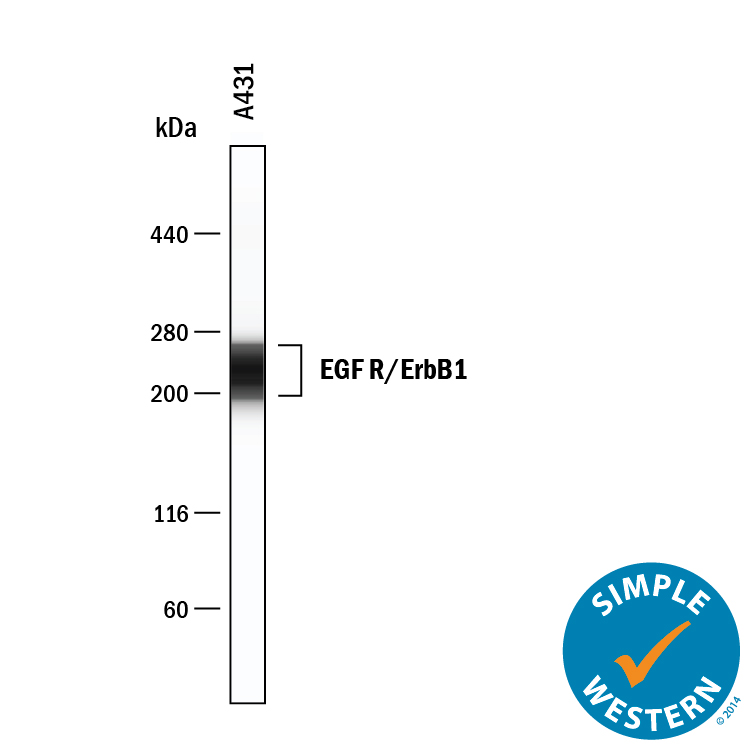
Detection of Human EGFR by Simple WesternTM. Simple Western lane view shows lysates of A431 human epithelial carcinoma cell line, loaded at 4.2 mg/mL. A specific band was detected for EGFR at approximately 229 kDa (as indicated) using 12.5 µg/mL of Goat Anti-Human EGFR Antigen Affinity-purified Polyclonal Antibody (Catalog # AF231). This experiment was conducted under reducing conditions and using the 12-230 kDa separation system.
Reconstitution Calculator
Preparation and Storage
- 12 months from date of receipt, -20 to -70 °C as supplied.
- 1 month, 2 to 8 °C under sterile conditions after reconstitution.
- 6 months, -20 to -70 °C under sterile conditions after reconstitution.
Background: EGFR
The epidermal growth factor receptor (EGFR) subfamily of receptor tyrosine kinases comprises four members: EGFR (also known as HER1, ErbB1 or ErbB), ErbB2 (Neu, HER2), ErbB3 (HER3), and ErbB4 (HER4). All family members are type I transmembrane glycoproteins that have an extracellular domain which contains two cysteine-rich domains separated by a spacer region that is involved in ligand binding, and a cytoplasmic domain which has a membrane-proximal tyrosine kinase domain and a C-terminal tail with multiple tyrosine autophosphorylation sites. The human EGFR gene encodes a 1210 amino acid (aa) residue precursor with a 24 aa putative signal peptide, a 621 aa extracellular domain, a 23 aa transmembrane domain, and a 542 aa cytoplasmic domain. EGFR has been shown to bind a subset of the EGF family ligands, including EGF, amphiregulin, TGF-alpha, betacellulin, epiregulin, heparin-binding EGF and neuregulin-2 alpha in the absence of a co-receptor. Ligand binding induces EGFR homodimerization as well as heterodimerization with ErbB2, resulting in kinase activation, tyrosine phosphorylation and cell signaling. EGFR can also be recruited to form heterodimers with the ligand-activated ErbB3 or ErbB4. EGFR signaling has been shown to regulate multiple biological functions including cell proliferation, differentiation, motility and apoptosis. In addition, EGFR signaling has also been shown to play a role in carcinogenesis (1 - 3).
- Daly, R.J. (1999) Growth Factors, 16:255.
- Schlessinger, J. (2000) Cell. 103:211.
- Maihle, N.J. et al. (2002) Cancer Treat. Res. 107:247.
Product Datasheets
Citations for Human EGFR Antibody
R&D Systems personnel manually curate a database that contains references using R&D Systems products. The data collected includes not only links to publications in PubMed, but also provides information about sample types, species, and experimental conditions.
16
Citations: Showing 1 - 10
Filter your results:
Filter by:
-
An atlas of late prenatal human neurodevelopment resolved by single-nucleus transcriptomics
Authors: SI Ramos, ZM Mussa, EN Falk, B Pai, B Giotti, K Allette, P Cai, F Dekio, R Sebra, KG Beaumont, AM Tsankov, NM Tsankova
Nature Communications, 2022-12-12;13(1):7671.
Species: Human
Sample Types: Whole Tissue
Applications: IHC/IF -
Plasma membrane proteoglycans syndecan-2 and syndecan-4 engage with EGFR and RON kinase to sustain carcinoma cell cycle progression
Authors: DM Beauvais, SE Nelson, KM Adams, NA Stueven, O Jung, AC Rapraeger
The Journal of Biological Chemistry, 2022-05-13;0(0):102029.
Species: Human
Sample Types: Cell Lysates
Applications: Immunoprecipitation, Western Blot -
Glioblastoma mutations alter EGFR dimer structure to prevent ligand bias
Authors: C Hu, CA Leche, A Kiyatkin, Z Yu, SE Stayrook, KM Ferguson, MA Lemmon
Nature, 2022-02-09;602(7897):518-522.
Species: Human
Sample Types: Cell Lysates
Applications: Western Blot -
Evaluation of the Targeting and Therapeutic Efficiency of Anti-EGFR Functionalised Nanoparticles in Head and Neck Cancer Cells for Use in NIR-II Optical Window
Authors: T Egnuni, N Ingram, I Mirza, PL Coletta, JR McLaughlan
Pharmaceutics, 2021-10-09;13(10):.
Species: Human
Sample Types: Whole Cell
Applications: IF -
Bacterial Antigens Reduced the Inhibition Effect of Capsaicin on Cal 27 Oral Cancer Cell Proliferation
Authors: R Chakrabort, K Vickery, C Darido, S Ranganatha, H Hu
International Journal of Molecular Sciences, 2021-08-12;22(16):.
Species: Human
Sample Types: Cell Lysates
Applications: Western Blot -
Involvement of cancer-derived EMT cells in the accumulation of 18F-fluorodeoxyglucose in the hypoxic cancer microenvironment
Authors: S Sugita, M Yamato, T Hatabu, Y Kataoka
Scientific Reports, 2021-05-17;11(1):9668.
Species: Mouse
Sample Types: Whole Tissue
Applications: IHC -
EGFR inhibition blocks cancer stem cell clustering and lung metastasis of triple negative breast cancer
Authors: X Liu, V Adorno-Cru, YF Chang, Y Jia, M Kawaguchi, NK Dashzeveg, R Taftaf, EK Ramos, EJ Schuster, L El-Shennaw, D Patel, Y Zhang, M Cristofani, H Liu
Theranostics, 2021-04-30;11(13):6632-6643.
Species: Human
Sample Types: Cell Lysates
Applications: Co-Immunoprecipitation -
Development of an immuno-wall device for the rapid and sensitive detection of EGFR mutations in tumor tissues resected from lung cancer patients
Authors: N Yogo, T Hase, T Kasama, K Nishiyama, N Ozawa, T Hatta, H Shibata, M Sato, K Komeda, N Kawabe, K Matsuoka, TF Chen-Yoshi, N Kaji, M Tokeshi, Y Baba, Y Hasegawa
PLoS ONE, 2020-11-16;15(11):e0241422.
Species: Human
Sample Types: Cell Lysates
Applications: ELISA Detection -
Identification of a novel anoikis signalling pathway using the fungal virulence factor gliotoxin
Authors: F Haun, S Neumann, L Peintner, K Wieland, J Habicht, C Schwan, K Østevold, MM Koczorowsk, M Biniossek, M Kist, H Busch, M Boerries, RJ Davis, U Maurer, O Schilling, K Aktories, C Borner
Nat Commun, 2018-08-30;9(1):3524.
Species: Human
Sample Types: Cell Lysates
Applications: Western Blot -
Improved efficiency of in situ protein analysis by proximity ligation using UnFold probes
Authors: A Klaesson, K Grannas, T Ebai, J Heldin, B Koos, M Leino, D Raykova, J Oelrich, L Arngården, O Söderberg, U Landegren
Sci Rep, 2018-03-29;8(1):5400.
Species: Human
Sample Types: Whole Tissue
Applications: IHC-P -
Soluble fms-like tyrosine kinase 1 promotes angiotensin II sensitivity in preeclampsia
Authors: Suzanne D Burke
J Clin Invest, 2016-06-06;0(0):.
Species: Human
Sample Types: Whole Tissue
Applications: IHC-P -
Ability of the Met kinase inhibitor crizotinib and new generation EGFR inhibitors to overcome resistance to EGFR inhibitors.
Authors: Nanjo, Shigeki, Yamada, Tadaaki, Nishihara, Hiroshi, Takeuchi, Shinji, Sano, Takako, Nakagawa, Takayuki, Ishikawa, Daisuke, Zhao, Lu, Ebi, Hiromich, Yasumoto, Kazuo, Matsumoto, Kunio, Yano, Seiji
PLoS ONE, 2013-12-26;8(12):e84700.
Species: Human
Sample Types: Cell Lysates
Applications: Western Blot -
Human epidermal growth factor receptor (HER-1:HER-3) Fc-mediated heterodimer has broad antiproliferative activity in vitro and in human tumor xenografts.
Authors: Sarup J, Jin P, Turin L, Bai X, Beryt M, Brdlik C, Higaki JN, Jorgensen B, Lau FW, Lindley P, Liu J, Ni I, Rozzelle J, Kumari R, Watson SA, Zhang J, Shepard HM
Mol. Cancer Ther., 2008-10-01;7(10):3223-36.
Species: Human
Sample Types: Cell Lysates, Recombinant Protein
Applications: ELISA Development, Western Blot -
Development and validation of sandwich ELISA microarrays with minimal assay interference.
Authors: Gonzalez RM, Seurynck-Servoss SL, Crowley SA
J. Proteome Res., 2008-04-19;7(6):2406-14.
Species: Human
Sample Types: Serum
Applications: ELISA Microarray Development -
FGFR2-amplified gastric cancer cell lines require FGFR2 and Erbb3 signaling for growth and survival.
Authors: Kunii K, Davis L, Gorenstein J, Hatch H, Yashiro M, Di Bacco A, Elbi C, Lutterbach B
Cancer Res., 2008-04-01;68(7):2340-8.
Species: Human
Sample Types: Cell Lysates
Applications: Immunoprecipitation, Western Blot -
Expression of growth factors and growth factor receptor in non-healing and healing ischaemic ulceration.
Authors: Murphy MO, Ghosh J, Fulford P, Khwaja N, Halka AT, Carter A, Turner NJ, Walker MG
Eur J Vasc Endovasc Surg, 2006-01-20;31(5):516-22.
Species: Human
Sample Types: Whole Tissue
Applications: IHC
FAQs
No product specific FAQs exist for this product, however you may
View all Antibody FAQsReviews for Human EGFR Antibody
Average Rating: 5 (Based on 1 Review)
Have you used Human EGFR Antibody?
Submit a review and receive an Amazon gift card.
$25/€18/£15/$25CAN/¥75 Yuan/¥2500 Yen for a review with an image
$10/€7/£6/$10 CAD/¥70 Yuan/¥1110 Yen for a review without an image
Filter by:


