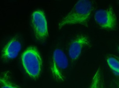
Human/Mouse/Rat Vimentin Antibody Summary
Ser2-Glu466
Accession # P08670
Applications
Please Note: Optimal dilutions should be determined by each laboratory for each application. General Protocols are available in the Technical Information section on our website.
Scientific Data
 View Larger
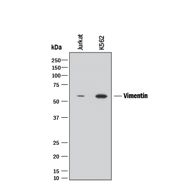
Detection of Human Vimentin by Western Blot. Western blot shows lysates of Jurkat human acute T cell leukemia cell line and K562 human chronic myelogenous leukemia cell line. PVDF membrane was probed with 2 µg/mL of Rat Anti-Human/Mouse/Rat Vimentin Monoclonal Antibody (Catalog # MAB2105) followed by HRP-conjugated Anti-Rat IgG Secondary Antibody (Catalog # HAF005). A specific band was detected for Vimentin at approximately 55 kDa (as indicated). This experiment was conducted under reducing conditions and using Immunoblot Buffer Group 1.
 View Larger
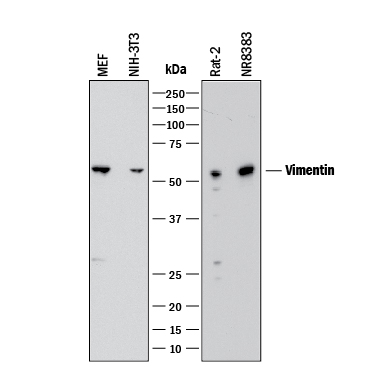
Detection of Mouse and Rat Vimentin by Western Blot. Western blot shows lysates of MEF mouse embryonic feeder cells, NIH-3T3 mouse embryonic fibroblast cell line, Rat-2 rat embryonic fibroblast cell line, and NR8383 rat alveolar macrophage cell line. PVDF membrane was probed with 1 µg/mL of Rat Anti-Human/Mouse/Rat Vimentin Monoclonal Antibody (Catalog # MAB2105) followed by HRP-conjugated Anti-Rat IgG Secondary Antibody (Catalog # HAF005). A specific band was detected for Vimentin at approximately 55 kDa (as indicated). This experiment was conducted under reducing conditions and using Immunoblot Buffer Group 1.
 View Larger
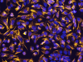
Vimentin in NTera‑2 Human Cell Line. Vimentin was detected in immersion fixed NTera-2 human testicular embryonic carcinoma cell line using Rat Anti-Human/Mouse/Rat Vimentin Monoclonal Antibody (Catalog # MAB2105) at 10 µg/mL for 3 hours at room temperature. Cells were stained using the NorthernLights™ 557-conjugated Anti-Rat IgG Secondary Antibody (yellow; Catalog # NL013) and counterstained with DAPI (blue). View our protocol for Fluorescent ICC Staining of Cells on Coverslips.
 View Larger
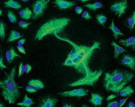
Vimentin in A549 Human Cell Line. Vimentin was detected in immersion fixed A549 human lung carcinoma cell line using Rat Anti-Human/Mouse/Rat Vimentin Monoclonal Antibody (Catalog # MAB2105) at 10 µg/mL for 3 hours at room temperature. Cells were stained using the NorthernLights™ 493-conjugated Anti-Rat IgG Secondary Antibody (green; Catalog # NL015) and counterstained with DAPI (blue). View our protocol for Fluorescent ICC Staining of Cells on Coverslips.
 View Larger
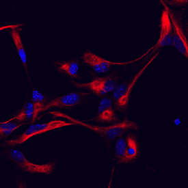
Vimentin in Mouse Cortical Stem Cells. Vimentin was detected in immersion fixed mouse cortical stem cells using Rat Anti-Human/Mouse/Rat Vimentin Monoclonal Antibody (Catalog # MAB2105) at 10 µg/mL for 3 hours at room temperature. Cells were stained using the NorthernLights™ 557-conjugated Anti-Rat IgG Secondary Antibody (red; Catalog # NL013) and counterstained with DAPI (blue). Specific staining was localized to cytoskeleton. View our protocol for Fluorescent ICC Staining of Cells on Coverslips.
 View Larger
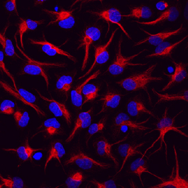
Vimentin in Rat Cortical Stem Cells. Vimentin was detected in immersion fixed rat cortical stem cells using Rat Anti-Human/Mouse/Rat Vimentin Monoclonal Antibody (Catalog # MAB2105) at 10 µg/mL for 3 hours at room temperature. Cells were stained using the NorthernLights™ 557-conjugated Anti-Rat IgG Secondary Antibody (red; Catalog # NL013) and counterstained with DAPI (blue). Specific staining was localized to cytoskeleton. View our protocol for Fluorescent ICC Staining of Cells on Coverslips.
 View Larger
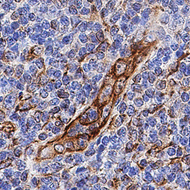
Vimentin in Human Tonsil. Vimentin was detected in immersion fixed paraffin-embedded sections of human tonsil using Rat Anti-Human/Mouse/Rat Vimentin Monoclonal Antibody (Catalog # MAB2105) at 0.5 µg/mL for 1 hour at room temperature followed by incubation with the Anti-Rat IgG VisUCyte™ HRP Polymer Antibody (Catalog # VC005). Tissue was stained using DAB (brown) and counterstained with hematoxylin (blue). Specific staining was localized to cytoplasm. View our protocol for IHC Staining with VisUCyte HRP Polymer Detection Reagents.
 View Larger
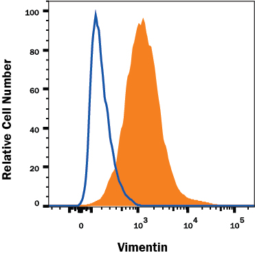
Detection of Vimentin in A172 Human Cell Line by Flow Cytometry. A172 human glioblastoma cell line was stained with Rat Anti-Human/Mouse/Rat Vimentin Monoclonal Antibody (Catalog # MAB2105, filled histogram) or isotype control antibody (Catalog # MAB006, open histogram) followed by anti-Rat IgG PE-conjugated secondary antibody (Catalog # F0105B). To facilitate intracellular staining, cells were fixed with Flow Cytometry Fixation Buffer (Catalog # FC004) and permeabilized with Flow Cytometry Permeabilization/Wash Buffer I (Catalog # FC005). View our protocol for Staining Intracellular Molecules.
 View Larger
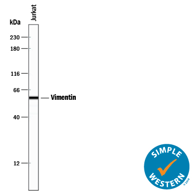
Detection of Human Vimentin by Simple WesternTM. Simple Western lane view shows lysates of Jurkat human acute T cell leukemia cell line, loaded at 0.2 mg/mL. A specific band was detected for Vimentin at approximately 58 kDa (as indicated) using 10 µg/mL of Rat Anti-Human/Mouse/Rat Vimentin Monoclonal Antibody (Catalog # MAB2105) followed by 1:50 dilution of HRP-conjugated Anti-Rat IgG Secondary Antibody (Catalog # HAF005). This experiment was conducted under reducing conditions and using the 12-230 kDa separation system.
 View Larger
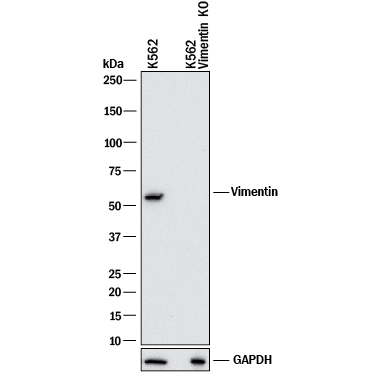
Western Blot Shows Human Vimentin Specificity by Using Knockout Cell Line. Western blot shows lysates of K562 human chronic myelogenous leukemia parental cell line and Vimentin knockout K562 cell line (KO). PVDF membrane was probed with 2 µg/mL of Rat Anti-Human/Mouse/Rat Vimentin Monoclonal Antibody (Catalog # MAB2105) followed by HRP-conjugated Anti-Goat IgG Secondary Antibody (HAF017). A specific band was detected for Vimentin at approximately 55 kDa (as indicated) in the parental K562 cell line, but is not detectable in knockout K562 cell line. GAPDH (MAB5718) is shown as a loading control. This experiment was conducted under reducing conditions and using Western Blot Buffer Group 1.
Reconstitution Calculator
Preparation and Storage
- 12 months from date of receipt, -20 to -70 °C as supplied.
- 1 month, 2 to 8 °C under sterile conditions after reconstitution.
- 6 months, -20 to -70 °C under sterile conditions after reconstitution.
Background: Vimentin
Vimentin is a 57 kDa class III intermediate filament (IF) protein that belongs to the intermediate filament family. It is the predominant IF in cells of mesenchymal origin such as vascular endothelium and blood cells (1-3). The human Vimentin cDNA encodes a 466 amino acid (aa) protein that contains head and tail regions with multiple regulatory Ser/Thr phosphorylation sites, and a central rod domain with three coiled-coil regions separated by linkers (1, 2). Human Vimentin shares 97-98% aa identity with mouse, rat, ovine, bovine, and canine Vimentin. Sixteen Vimentin coiled-coil dimers self-assemble to form intermediate (10-12 nm wide) filaments (4). These filaments then anneal longitudinally to form non-polarized fibers that support cell structure and withstand stress (4). IF fibers are highly dynamic, and half-life depends on the balance between kinase and phosphatase activity. For example, phosphorylation followed by dephosphorylation drives IF disintegration, followed by reorganization during mitosis (1, 5, 6). Interactions of head and tail domains link IFs with other structures such as actin and microtubule cytoskeletons (7). Vimentin is involved in positioning autophagosomes, lysosomes and the Golgi complex within the cell (8). It facilitates cell migration and motility by recycling internalized trailing edge integrins back to the cell surface at the leading edge (9-11). Vimentin helps maintain the lipid composition of cellular membranes, and caspase cleavage of Vimentin is a key event in apoptosis (8, 12). Phosphorylation promotes secretion of Vimentin by TNF-alpha -stimulated macrophages (13). Extracellular Vimentin has been shown to associate with several microbes, and appears to promote an antimicrobial oxidative burst (13, 14). Cell-associated Vimentin can also interact with NKp46 to recruit NK cells to tuberculosis-infected monocytes (15).
- Omary, M.B. et al. (2006) Trends Biochem. Sci. 31:383.
- Ivaska, J. et al. (2007) Exp. Cell Res. 313:2050.
- Ferrari, S. et al. (1986) Mol. Cell. Biol. 6:3614.
- Sokolova, A.V. et al. (2006) Proc. Natl. Acad. Sci. USA 103:16206.
- Eriksson, J.E. et al. (2004) J. Cell Sci. 117:919.
- Li, Q-F. et al. (2006) J. Biol. Chem. 281:34716.
- Esue, O. et al. (2006) J. Biol. Chem. 281:30393.
- Styers, M.L. et al. (2005) Traffic 6:359.
- McInroy, L. and A. Maata (2007) Biochem. Biophys. Res. Commun. 360:109.
- Nieminen, M. et al. (2006) Nat. Cell Biol. 8:156.
- Ivaska, J. et al. (2005) EMBO J. 24:3834.
- Byun, Y. et al. (2001) Cell Death Differ. 8:443.
- Mor-Vaknin, N. et al. (2003) Nat. Cell Biol. 5:59.
- Zou, Y. et al. (2006) Biochem. Biophys. Res. Commun. 351:625.
- Garg, A. et al. (2006) J. Immunol. 177:6192.
Product Datasheets
Citations for Human/Mouse/Rat Vimentin Antibody
R&D Systems personnel manually curate a database that contains references using R&D Systems products. The data collected includes not only links to publications in PubMed, but also provides information about sample types, species, and experimental conditions.
21
Citations: Showing 1 - 10
Filter your results:
Filter by:
-
Transplantation of adipose tissue-derived microvascular fragments promotes therapy of critical limb ischemia
Authors: Park, GT;Lim, JK;Choi, EB;Lim, MJ;Yun, BY;Kim, DK;Yoon, JW;Hong, YG;Chang, JH;Bae, SH;Ahn, JY;Kim, JH;
Biomaterials research
Species: Human hepegivirus
Sample Types: Whole Cells
Applications: Flow Cytometry -
T cell deficiency precipitates antibody evasion and emergence of neurovirulent polyomavirus
Authors: MD Lauver, G Jin, KN Ayers, SN Carey, CS Specht, CS Abendroth, AE Lukacher
Elife, 2022-11-07;11(0):.
Species: Mouse
Sample Types: Whole Tissue
Applications: IHC -
Engineering of immune checkpoints B7-H3 and CD155 enhances immune compatibility of MHC-I-/- iPSCs for beta cell replacement
Authors: R Chimienti, T Baccega, S Torchio, F Manenti, S Pellegrini, A Cospito, A Amabile, MT Lombardo, P Monti, V Sordi, A Lombardo, M Malnati, L Piemonti
Cell Reports, 2022-09-27;40(13):111423.
Species: Human
Sample Types: Whole Cells
Applications: ICC -
NR2F1 is a barrier to dissemination of early stage breast cancer cells
Authors: C Rodriguez-, N Kale, MJ Carlini, N Shrivastav, AA Rodrigues, B Khalil, JJ Bravo-Cord, M Alexander, J Ji, Y Hong, F Behbod, MS Sosa
Cancer Research, 2022-06-15;0(0):.
Species: Mouse
Sample Types: Whole Cells
Applications: IF -
Suspension culture promotes serosal mesothelial development in human intestinal organoids
Authors: MM Capeling, S Huang, CJ Childs, JH Wu, YH Tsai, A Wu, N Garg, EM Holloway, N Sundaram, C Bouffi, M Helmrath, JR Spence
Cell Reports, 2022-02-15;38(7):110379.
Species: Human
Sample Types: Organoids
Applications: IHC -
The LRRK2 G2019S mutation alters astrocyte-to-neuron communication via extracellular vesicles and induces neuron atrophy in a human iPSC-derived model of Parkinson's disease.
Authors: de Rus Jacquet A, Tancredi J, Lemire A, DeSantis M, Li W, O'Shea E
Elife, 2021-09-30;10(0):.
Species: Human
Sample Types: Whole Cells
Applications: ICC -
Nrf1 promotes heart regeneration and repair by regulating proteostasis and redox balance
Authors: M Cui, A Atmanli, MG Morales, W Tan, K Chen, X Xiao, L Xu, N Liu, R Bassel-Dub, EN Olson
Nature Communications, 2021-09-06;12(1):5270.
Species: Mouse
Sample Types: Whole Tissue
Applications: IHC -
Echinochrome A Treatment Alleviates Fibrosis and Inflammation in Bleomycin-Induced Scleroderma
Authors: GT Park, JW Yoon, SB Yoo, YC Song, P Song, HK Kim, J Han, SJ Bae, KT Ha, NP Mishchenko, SA Fedoreyev, VA Stonik, MB Kim, JH Kim
Marine Drugs, 2021-04-23;19(5):.
Species: Mouse
Sample Types: Whole Tissue
Applications: IHC -
Primary cilia-dependent lipid raft/caveolin dynamics regulate adipogenesis
Authors: D Yamakawa, D Katoh, K Kasahara, T Shiromizu, M Matsuyama, C Matsuda, Y Maeno, M Watanabe, Y Nishimura, M Inagaki
Cell Reports, 2021-03-09;34(10):108817.
Species: Mouse
Sample Types: Whole Cells, Whole Tissue
Applications: ICC, IHC -
Heterogeneous Manifestations of Epithelial-Mesenchymal Plasticity of Circulating Tumor Cells in Breast Cancer Patients
Authors: LA Tashireva, OE Savelieva, ES Grigoryeva, YV Nikitin, EV Denisov, SV Vtorushin, MV Zavyalova, NV Cherdyntse, VM Perelmuter
International Journal of Molecular Sciences, 2021-03-02;22(5):.
Species: Human
Sample Types: Whole Cells
Applications: Flow Cytometry -
Induced organoids derived from patients with ulcerative colitis recapitulate colitic reactivity
Authors: SK Sarvestani, S Signs, B Hu, Y Yeu, H Feng, Y Ni, DR Hill, RC Fisher, S Ferrandon, RK DeHaan, J Stiene, M Cruise, TH Hwang, X Shen, JR Spence, EH Huang
Nature Communications, 2021-01-11;12(1):262.
Species: Human
Sample Types: Organoid
Applications: IHC -
Cell-Type-Specific Gene Regulatory Networks Underlying Murine Neonatal Heart Regeneration at Single-Cell Resolution
Authors: Z Wang, M Cui, AM Shah, W Tan, N Liu, R Bassel-Dub, EN Olson
Cell Reports, 2020-12-08;33(10):108472.
Species: Human
Sample Types: Whole Cells
Applications: ICC -
Quantitative proteomic comparison of myofibroblasts derived from bone marrow and cornea
Authors: P Saikia, JS Crabb, LL Dibbin, MJ Juszczak, B Willard, GF Jang, TM Shiju, JW Crabb, SE Wilson
Sci Rep, 2020-10-07;10(1):16717.
Species: Rabbit
Sample Types: Whole Cells
Applications: ICC -
Sensitive and easy screening for circulating tumor cells by flow cytometry
Authors: A Lopresti, F Malergue, F Bertucci, ML Liberatosc, S Garnier, Q DaCosta, P Finetti, M Gilabert, JL Raoul, D Birnbaum, C Acquaviva, E Mamessier
JCI Insight, 2019-06-13;5(0):.
Species: Human
Sample Types: Whole Cells
Applications: Flow Cytometry -
Platelet glycoprotein VI and C-type lectin-like receptor 2 deficiency accelerates wound healing by impairing vascular integrity in mice
Authors: S Wichaiyo, S Lax, SJ Montague, Z Li, B Grygielska, JA Pike, EJ Haining, A Brill, SP Watson, J Rayes
Haematologica, 2019-02-07;0(0):.
Species: Mouse
Sample Types: Whole Tissue
Applications: IHC-P -
RNA sequencing reveals upregulation of a transcriptomic program associated with stemness in metastatic prostate cancer cells selected for taxane resistance
Authors: CK Cajigas-Du, SR Martinez, L Woods-Burn, AM Durán, S Roy, A Basu, JA Ramirez, GL Ortiz-Hern, L Ríos-Colón, E Chirshev, ES Sanchez-He, U Soto, C Greco, C Boucheix, X Chen, J Unternaehr, C Wang, CA Casiano
Oncotarget, 2018-07-13;9(54):30363-30384.
Species: Human
Sample Types: Cell Lysates
Applications: Western Blot -
Acute Drug Effects on the Human Placental Tissue: The Development of a Placental Murine Xenograft Model
Authors: M Verheecke, E Hermans, S Tuyaerts, E Souche, R Van Bree, G Verbist, T Everaert, J Van Houdt, K Van Calste, F Amant
Reprod Sci, 2018-02-13;0(0):1933719118756.
Species: Xenograft
Sample Types: Whole Cells
Applications: Flow Cytometry -
Combined CSL and p53 downregulation promotes cancer-associated fibroblast activation.
Authors: Procopio M, Laszlo C, Al Labban D, Kim D, Bordignon P, Jo S, Goruppi S, Menietti E, Ostano P, Ala U, Provero P, Hoetzenecker W, Neel V, Kilarski W, Swartz M, Brisken C, Lefort K, Dotto G
Nat Cell Biol, 2015-08-24;17(9):1193-204.
Species: Human
Sample Types: Whole Tissue
Applications: IHC -
The TNF Family Molecules LIGHT and Lymphotoxin alphabeta Induce a Distinct Steroid-Resistant Inflammatory Phenotype in Human Lung Epithelial Cells.
Authors: da Silva Antunes R, Madge L, Soroosh P, Tocker J, Croft M
J Immunol, 2015-07-24;195(5):2429-41.
-
GLI1, CTNNB1 and NOTCH1 protein expression in a thymic epithelial malignancy tissue microarray.
Authors: Riess J, West R, Dean M, Klimowicz A, Neal J, Hoang C, Wakelee H
2015-02-01;0(0):.
Species: Human
Sample Types: Whole Tissue
Applications: IHC -
Development of a reconstructed cornea from collagen-chondroitin sulfate foams and human cell cultures.
Authors: Vrana NE, Builles N, Justin V, Bednarz J, Pellegrini G, Ferrari B, Damour O, Hulmes DJ, Hasirci V
Invest. Ophthalmol. Vis. Sci., 2008-08-15;49(12):5325-31.
Species: Human
Sample Types: Whole Tissue
Applications: IHC-P
FAQs
No product specific FAQs exist for this product, however you may
View all Antibody FAQsReviews for Human/Mouse/Rat Vimentin Antibody
Average Rating: 4.4 (Based on 5 Reviews)
Have you used Human/Mouse/Rat Vimentin Antibody?
Submit a review and receive an Amazon gift card.
$25/€18/£15/$25CAN/¥75 Yuan/¥2500 Yen for a review with an image
$10/€7/£6/$10 CAD/¥70 Yuan/¥1110 Yen for a review without an image
Filter by:
Vimentin expression in human fibroblasts in culture. Vimentin (green) was used as a fibroblast marker in miofibroblast differentiation assays (a-SMA in red, as a miofibroblast marker).
