Human/Rat GFAP Antibody Summary
Leu292-Met432
Accession # P14136
Applications
Please Note: Optimal dilutions should be determined by each laboratory for each application. General Protocols are available in the Technical Information section on our website.
Scientific Data
 View Larger
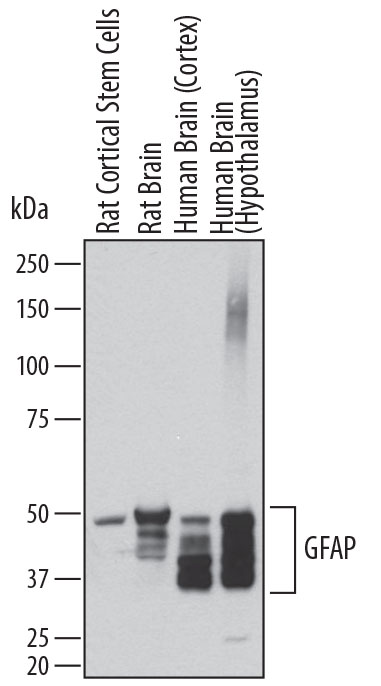
Detection of Human and Rat GFAP by Western Blot. Western blot shows lysates of rat cortical stem cells, rat brain tissue, human brain (cortex) tissue, and human brain (hypothalamus) tissue. PVDF membrane was probed with 0.2 µg/mL of Sheep Anti-Human GFAP Antigen Affinity-purified Polyclonal Antibody (Catalog # AF2594) followed by HRP-conjugated Anti-Sheep IgG Secondary Antibody (Catalog # HAF016). Specific bands were detected for GFAP at approximately 35-50 kDa (as indicated). This experiment was conducted under reducing conditions and using Immunoblot Buffer Group 1.
 View Larger
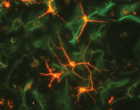
beta ‑III Tubulin in Rat Cortical Neurons and GFAP in Rat Astrocytes. beta -III Tubulin was detected in rat cortical neurons using 5 µg/mL Mouse Anti-neuron-specific Mouse beta -III Tubulin Monoclonal (clone TuJ-1) Antibody (Catalog # MAB1195). GFAP was detected in rat astrocytes using 10 µg/mL Sheep Anti-Human GFAP Antigen Affinity-purified Poly-clonal Antibody (Catalog # AF2594). Cells were incubated with primary antibodies for 3 hours at room temperature. Cells were stained for beta-III Tubulin using the Northern-Lights™ 557-conjugated Anti-Mouse IgG Secondary Antibody (red; Catalog # NL007) and for GFAP using the Northern-Lights 493-conjugated Anti-Sheep IgG Secondary Antibody (green; Catalog # NL012). View our protocol for Fluorescent ICC Staining of Cells on Coverslips.
 View Larger
GFAP in Rat Astrocytes. GFAP was detected in immersion fixed rat astrocytes using 10 µg/mL Sheep Anti-Human GFAP Antigen Affinity-purified Polyclonal Antibody (Catalog # AF2594) for 3 hours at room temperature. Cells were stained with the NorthernLights™ 557-conjugated Anti-Sheep IgG Secondary Antibody (red; Catalog # NL010) and counterstained with DAPI (blue). View our protocol for Fluorescent ICC Staining of Cells on Coverslips.
 View Larger
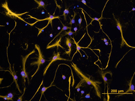
GFAP in Rat Cortical Stem Cells. GFAP was detected in immersion fixed 7 days differentiated rat cortical stem cells using Sheep Anti-Human GFAP Antigen Affinity-purified Poly-clonal Antibody (Catalog # AF2594) at 10 µg/mL for 3 hours at room temperature. Cells were stained using the Northern-Lights™ 557-conjugated Anti-Sheep IgG Secondary Antibody (yellow; Catalog # NL010) and counterstained with DAPI (blue). View our protocol for Fluorescent ICC Staining of Cells on Coverslips.
 View Larger
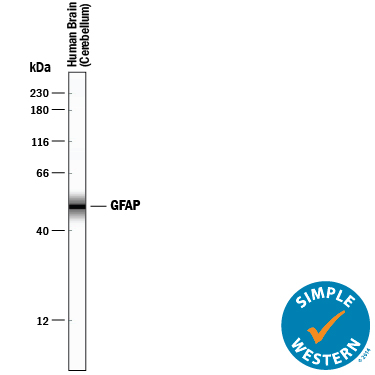
Detection of Human GFAP by Simple WesternTM. Simple Western lane view shows lysates of human brain (cerebellum) tissue, loaded at 0.2 mg/mL. A specific band was detected for GFAP at approximately 51 kDa (as indicated) using 0.1 µg/mL of Sheep Anti-Human/Rat GFAP Antigen Affinity-purified Polyclonal Antibody (Catalog # AF2594) followed by 1:50 dilution of HRP-conjugated Anti-Sheep IgG Secondary Antibody (Catalog # HAF016). This experiment was conducted under reducing conditions and using the 12-230 kDa separation system.
 View Larger
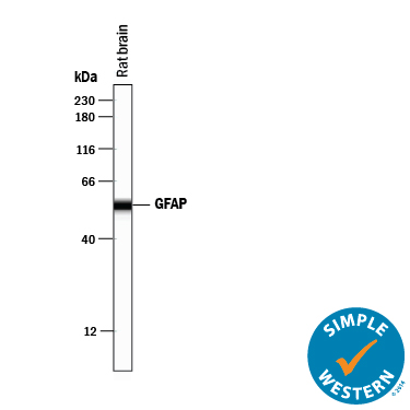
Detection of Rat GFAP by Simple WesternTM. Simple Western lane view shows lysates of rat brain tissue, loaded at 0.2 mg/mL. A specific band was detected for GFAP at approximately 55 kDa (as indicated) using 2 µg/mL of Sheep Anti-Human/Rat GFAP Antigen Affinity-purified Polyclonal Antibody (Catalog # AF2594) followed by 1:50 dilution of HRP-conjugated Anti-Sheep IgG Secondary Antibody (Catalog # HAF016). This experiment was conducted under reducing conditions and using the 12-230 kDa separation system.
Reconstitution Calculator
Preparation and Storage
- 12 months from date of receipt, -20 to -70 °C as supplied.
- 1 month, 2 to 8 °C under sterile conditions after reconstitution.
- 6 months, -20 to -70 °C under sterile conditions after reconstitution.
Background: GFAP
GFAP (Glial fibrillary acidic protein) is a type III intermediate filament protein. It is the major component of astrocyte intermediate filament. Defects in GFAP are a cause of Alexander disease. Alexander disease is a rare disorder of the central nervous system. It is a progressive leukoencephalopathy whose hallmark is the widespread accumulation of Rosenthal fibers which are cytoplasmic inclusions in astrocytes. At the amino acid sequence level, human GFAP shares 91% and 90% identity with rat and mouse GFAP, respectively.
Product Datasheets
Citations for Human/Rat GFAP Antibody
R&D Systems personnel manually curate a database that contains references using R&D Systems products. The data collected includes not only links to publications in PubMed, but also provides information about sample types, species, and experimental conditions.
7
Citations: Showing 1 - 7
Filter your results:
Filter by:
-
A single-cell transcriptome atlas of glial diversity in the human hippocampus across the postnatal lifespan
Authors: Y Su, Y Zhou, ML Bennett, S Li, M Carceles-C, L Lu, S Huh, D Jimenez-Cy, BC Kennedy, SK Kessler, AN Viaene, I Helbig, X Gu, JE Kleinman, TM Hyde, DR Weinberger, DW Nauen, H Song, GL Ming
Cell Stem Cell, 2022-11-03;29(11):1594-1610.e8.
Species: Human
Sample Types: Whole Tissue
Applications: IHC -
Anti-M�llerian Hormone Regulation of Synaptic Transmission in the Hippocampus Requires MAPK Signaling and Kv4.2 Potassium Channel Activity
Authors: K Wang, F Xu, J Maylie, J Xu
Frontiers in Neuroscience, 2021-12-16;15(0):772251.
Species: Mouse
Sample Types: Whole Tissue
Applications: IHC -
Modeling the mature CNS: A predictive screening platform for neurodegenerative disease drug discovery
Authors: KM LaBarbera, C Limegrover, C Rehak, R Yurko, NJ Izzo, N Knezovich, E Watto, L Waybright, SM Catalano
Journal of neuroscience methods, 2021-04-06;0(0):109180.
Species: Human
Sample Types: Whole Cells
Applications: ICC -
APP upregulation contributes to retinal ganglion cell degeneration via JNK3
Authors: C Liu, CW Zhang, Y Zhou, WQ Wong, LC Lee, WY Ong, SO Yoon, W Hong, XY Fu, TW Soong, EH Koo, LW Stanton, KL Lim, ZC Xiao, GS Dawe
Cell Death Differ., 2017-12-13;0(0):.
Species: Mouse
Sample Types: Whole Tissue
Applications: IHC-P -
Time-Dependent, HIV-Tat-Induced Perturbation of Human Neurons In Vitro: Towards a Model for the Molecular Pathology of HIV-Associated Neurocognitive Disorders
Authors: KT Gurwitz, RJ Burman, BD Murugan, S Garnett, T Ganief, NC Soares, JV Raimondo, JM Blackburn
Front Mol Neurosci, 2017-05-29;10(0):163.
Species: Human
Sample Types: Whole Cells
Applications: ICC -
Dose-dependent changes in neuroinflammatory and arachidonic acid cascade markers with synaptic marker loss in rat lipopolysaccharide infusion model of neuroinflammation.
BMC Neurosci, 2012-05-23;13(0):50.
Species: Rat
Sample Types: Tissue Homogenates
Applications: Western Blot -
TNFalpha-induced AMPA-receptor trafficking in CNS neurons; relevance to excitotoxicity?
Authors: Leonoudakis D, Braithwaite SP, Beattie MS, Beattie EC
Neuron Glia Biol., 2004-08-01;1(3):263-273.
Species: Rat
Sample Types: Whole Cells
Applications: ICC
FAQs
No product specific FAQs exist for this product, however you may
View all Antibody FAQsReviews for Human/Rat GFAP Antibody
There are currently no reviews for this product. Be the first to review Human/Rat GFAP Antibody and earn rewards!
Have you used Human/Rat GFAP Antibody?
Submit a review and receive an Amazon gift card.
$25/€18/£15/$25CAN/¥75 Yuan/¥2500 Yen for a review with an image
$10/€7/£6/$10 CAD/¥70 Yuan/¥1110 Yen for a review without an image


