Human SCD-1 Antibody Summary
Applications
Please Note: Optimal dilutions should be determined by each laboratory for each application. General Protocols are available in the Technical Information section on our website.
Scientific Data
 View Larger
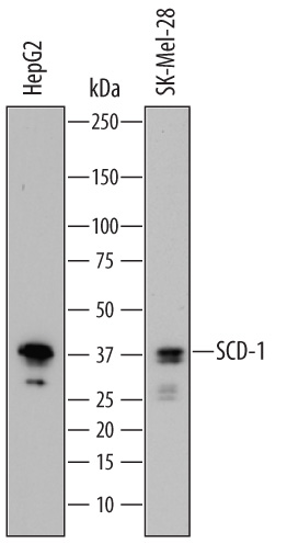
Detection of Human SCD‑1 by Western Blot. Western blot shows lysates of HepG2 human hepatocellular carcinoma cell line and SK-Mel-28 human malignant melanoma cell line. PVDF membrane was probed with 0.5 µg/mL of Sheep Anti-Human SCD-1 Antigen Affinity-purified Polyclonal Antibody (Catalog # AF7550) followed by HRP-conjugated Anti-Sheep IgG Secondary Antibody (Catalog # HAF016). A specific band was detected for SCD-1 at approximately 38 kDa (as indicated). This experiment was conducted under reducing conditions and using Immunoblot Buffer Group 1.
 View Larger
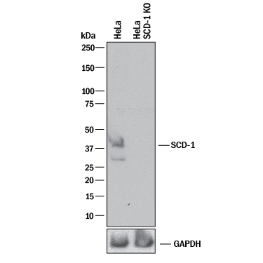
Western Blot Shows Human SCD‑1 Specificity by Using Knockout Cell Line. Western blot shows lysates of HeLa human cervical epithelial carcinoma parental cell line and SCD-1 knockout HeLa cell line (KO). PVDF membrane was probed with 0.5 µg/mL of Sheep Anti-Human SCD-1 Antigen Affinity-purified Polyclonal Antibody (Catalog # AF7550) followed by HRP-conjugated Anti-Sheep IgG Secondary Antibody (Catalog # HAF016). A specific band was detected for SCD-1 at approximately 41 kDa (as indicated) in the parental HeLa cell line, but is not detectable in knockout HeLa cell line. GAPDH (Catalog # AF5718) is shown as a loading control. This experiment was conducted under reducing conditions and using Immunoblot Buffer Group 1.
Reconstitution Calculator
Preparation and Storage
- 12 months from date of receipt, -20 to -70 degreesC as supplied. 1 month, 2 to 8 degreesC under sterile conditions after reconstitution. 6 months, -20 to -70 degreesC under sterile conditions after reconstitution.
Background: SCD-1
SCD-1 (Stearoyl-CoA desaturase 1; also Acyl-CoA desaturase, fatty acid desaturase, and Delta-9 desaturase) is a 37-40 kDa member of the fatty acid desaturase family of enzymes. It is an ER-embedded protein that is expressed by multiple cell types, including adipocytes, hepatocytes, macrophages, endothelial and sebaceous gland cells. SCD-1 catalyzes the formation of monounsaturated fatty acids from saturated fatty acids. It does so by generating a double bond between the C9 and C10 carbons of dietary and/or endogenously synthesized fatty acids. This creates either palmitoleic or oleic acid, two fatty acids that are optimally suited for either storage or inclusion into phospholipids. It also removes a potential source of inflammation, as saturated fatty acids are known to activate TLRs with the subsequent onset of inflammation. Human SCD-1 is a 4-transmembrane (TM), 359 amino acid (aa) protein. It contains a 71 aa cytoplasmic N-terminus, followed by two TM segments (aa 72-119) and an extended cytoplasmic region (aa 120-216) that possesses three utilized Ser/Thr phosphorylation sites, two additional TM segments (aa 217-273), and a C-terminal cytoplasmic tail (aa 274-359) that contains most of the catalytic region. There is one potential isoform variant that shows a 13 aa substitution for aa 295-359. Over aa 141-221, human SCD-1 shares 95% aa sequence identity with mouse SCD-1.
Product Datasheets
FAQs
No product specific FAQs exist for this product, however you may
View all Antibody FAQsReviews for Human SCD-1 Antibody
There are currently no reviews for this product. Be the first to review Human SCD-1 Antibody and earn rewards!
Have you used Human SCD-1 Antibody?
Submit a review and receive an Amazon gift card.
$25/€18/£15/$25CAN/¥75 Yuan/¥2500 Yen for a review with an image
$10/€7/£6/$10 CAD/¥70 Yuan/¥1110 Yen for a review without an image

