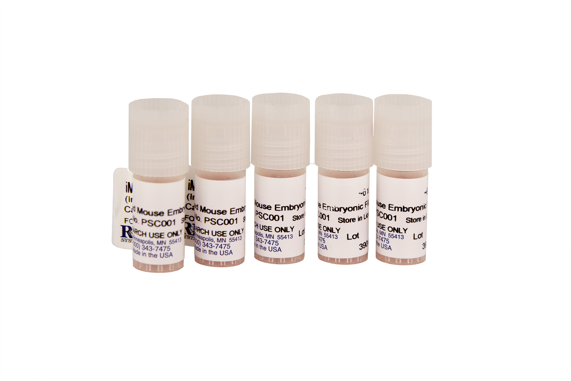
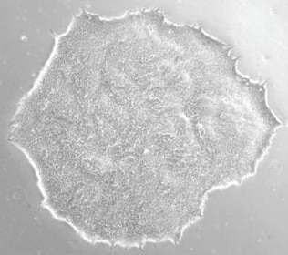
Irradiated Mouse Embryonic Fibroblasts (5 vials)
Irradiated Mouse Embryonic Fibroblasts (5 vials) Summary
For use as a feeder layer when culturing embryonic or induced pluripotent stem cells.
Key Benefits
- Ready to use
- MEFs support undifferentiated growth of embryonic and induced pluripotent stem cells
- Inactivated by gamma irradiation to avoid chemical contamination
- Consistent feeder layer to reduce experimental variation
- Free of microbial and mycoplasma contamination
The Importance of Mouse Embryonic Fibroblasts in Maintaining Undifferentiated Cells in Culture
Mitotically inactive mouse embryonic fibroblasts (MEFs) support the growth and maintenance of stem cell pluripotency by secreting necessary soluble factors and by providing a substrate for stem cell attachment.
However, the multi-step procedure used to generate mitotically inactive MEFs involves several opportunities for the introduction of experimental variation and contamination. MEFs of low quality and/or purity can compromise the growth, pluripotency, and viability of stem cells in culture. R&D Systems offers high quality MEFs that are mitotically arrested by irradiation at passage 3 and are free of microbial and mycoplasma contamination.
Five vials of irradiated mouse embryonic fibroblasts are provided with 6 x 10⁶ cells in each vial.
Mouse embryonic fibroblasts (MEF) were isolated from E13.5 CF1 embryos after removal of brain and visceral tissue. The MEF were then mitotically inactivated at passage 3 by gamma irradiation and cryopreserved.
Stability and Storage
Store in liquid nitrogen for up to 2 years.
Quality Control
R&D Systems iMEF are tested for their ability to support undifferentiated growth of BG01V human ES cells as assessed by Oct-4 and SSEA-4 expression. They are mycoplasma negative as tested by the MycoProbe™ Mycoplasma Detection Kit (Catalog # CUL001B) and negative for microbial contamination.
Precautions
This product contains 10% dimethylsulfoxide (DMSO).
Specifications
Product Datasheets
Scientific Data
 View Larger
Human Pluripotent Stem Cells Cultured on Mouse Embryonic Fibroblasts Express Pluripotent Markers SSEA-4, Oct-3/4, Oct-4A, and E-Cadherin. Human iPS2 stem cells were cultured on Irradiated Mouse Embryonic Fibroblasts (iMEF; Catalog # PSC001). Expression of SSEA-4 was detected using a Mouse Anti-Human/Mouse SSEA-4 Monoclonal Antibody (Catalog # MAB1435) followed by the NorthernLights™ (NL) 493-conjugated Donkey Anti-Mouse IgG Secondary Antibody (Catalog # NL009, green). Oct-3/4 expression was detected using a Goat Anti-Human Oct-3/4 Affinity Purified Polyclonal Antibody (Catalog # AF1759) followed by the NL557-conjugated Donkey Anti-Goat IgG (Catalog # NL001, red). The nuclei were counterstained with DAPI (blue).B.BG01V human embryonic stem cells were cultured on Irradiated Mouse Embryonic Fibroblasts (iMEF; Catalog # PSC001). Expression of Oct-4A was detected using a Mouse Anti-human Oct-4A Monoclonal Antibody (Catalog # MAB17591) followed by the NL493-conjugated Donkey Anti-mouse IgG Secondary Antibody (Catalog # NL009, green). Expression of E-Cadherin was detected using a Goat Anti-Human E-Cadherin Affinity Purified Polyclonal Antibody (Catalog # AF648) followed by the NL 557-conjugated Donkey Anti-Goat IgG (Catalog # NL001, red). Nuclei were counterstained with DAPI (blue). View our protocol for Fluorescent ICC Staining of Stem Cells on Coverslips.
 View Larger
BG01V Human Embryonic Stem Cells Cultured on Irradiated Mouse Embryonic Fibroblasts Demonstrate a Phenotype that is Consitent with Pluripotency. A. BG01V human embryonic stem cell colonies cultured on Irradiated Mouse Embryonic Fibroblasts (Catalog # PSC001) were analyzed for markers of pluripotency after 3 passages. B. Cells were evaluated for SSEA-4 expression by flow cytometry using a PE-conjugated Mouse Anti-Human SSEA-4 Monoclonal Antibody (Catalog # FAB1435P; filled histogram) or a PE-conjugated Mouse IgG3 Isotype Control Antibody (Catalog # IC007P; open histogram). C.&D. BG01V human embryonic stem cell colonies were incubated with a Goat Anti-Human Oct-3/4 Antigen Affinity-purified Polyclonal Antibody (Catalog # AF1759) or a Goat Anti-Human Nanog Antigen Affinity-purified Polyclonal Antibody (Catalog # AF1997). The cells were stained with the NorthernLights™ 557-conjugated Donkey Anti-Goat IgG Secondary Antibody (Catalog # NL001). Expression of SSEA-4, Oct-3/4, and Nanog is consistent with a pluripotent phenotype. View our protocol for Fluorescent ICC Staining of Stem Cells on Coverslips.
Assay Procedure
Refer to the product datasheet for complete product details.
Briefly, Irradiated Mouse Embryonic Fibroblasts (iMEFs) can be used as a feeder layer for pluripotent stem cells using the following procedure:
- Thaw iMEFs
- Plate iMEFs onto gelatin-coated plates and culture for 24 hours
- Plate and culture human pluripotent stem cells on iMEF-containing plates
Reagents Supplied in the Irradiated Mouse Embryonic Fibroblasts (Catalog # PSC001):
- 5 vials of irradiated mouse embryonic fibroblasts with 6 x 10⁶ cells/vial
Reagents
- BG01V human embryonic stem cells
- Fetal bovine serum
- Knockout™ serum replacer (Invitrogen)
- Non-Essential Amino Acids (100X)
- L-Glutamine (200 mM)
- Penicillin/Streptomycin (100X)
- beta-mercaptoethanol
- DMEM/F12
- High glucose DMEM
- Recombinant Human FGF basic (Catalog # 233-FB; or Tissue Culture Grade, Catalog # 4114-TC)
- Accutase® (Innovative Cell Technologies)
- 0.1% w/v solution of gelatin in sterile deionized H2O
Materials
- Tissue culture plates (60 mm; the protocol can be adapted for other plate sizes)
- 15 mL centrifuge tubes
- 0.2 µm sterile filter unit
- Pipettes and pipette tips
Equipment
- 37 °C and 5% CO2 incubator
- Centrifuge (low speed clinical or equivalent)
- Hemocytometer
- Microscope
Reagent and Media Preparation
Mouse Embryonic Fibroblast (MEF) Media - MEF media consists of high glucose DMEM, 10% fetal bovine serum, 2 mM L-glutamine, and if desired, add a 1:100 dilution of penicillin/streptomycin (100X) stock. Filter sterilize the media using 0.2 µm sterile filter unit.
Human Embryonic Stem (hES) Media – human embryonic stem cell media consists of DMEM/F12, 15% fetal bovine serum, 5% Knockout serum replacer, 1:100 dilution of non-essential amino acids stock (100X), 1:100 dilution of penicillin/streptomycin (100X) stock, and 0.1 µM beta-mercaptoethanol. Filter sterilize the media using 0.2 µm sterile filter unit. Just prior to use, the media should be supplemented with Recombinant Human FGF basic at 4 ng/mL.
I. Thawing and Plating of iMEF Feeder Cells (plate feeder cells one day prior to stem cell seeding)
Coat the appropriate sized plate(s) with 0.1% sterile Gelatin for 15 minutes.
Aspirate 0.1% Gelatin from plates.

Transfer thawed iMEFs to a 15 mL conical tube that contains 5 mL of pre-warmed Mouse Embryonic Fibroblast (MEF) media.
Centrifuge at 200 x g for 5 minutes.
Remove the supernatant and gently flick the pellet.
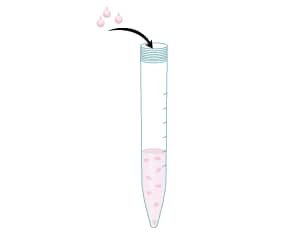
Resuspend the iMEF cells in MEF media.
Transfer the cells to the Gelatin-coated plates at a density of approximately 1 x 10⁶ cells/60 mm plate.
Incubate overnight at 37 °C and 5% CO2 incubator before seeding stem cells.

II. Day 2: Thawing and Plating of BG01V Human Embryonic Stem Cells
Transfer thawed BG01V human embryonic stem cells to a 15 mL centrifuge tube that contains at least 5 mL of pre-warmed hES media.
Centrifuge at 200 x g in a clinical centrifuge for 4 minutes.
Remove the supernatant and resuspend the pellet in hES Media supplemented with 4 ng/mL FGF basic.

Remove the MEF media from a plate containing iMEF cells.
Add the BG01V human embryonic stem cell suspension to the plate.
Incubate the cells at 37 °C and 5% CO2.
Replace media daily.
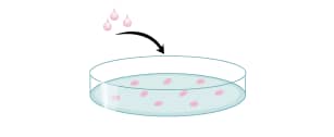
Remove the hES media from BG01V human embryonic stem cells.
Add 1 mL of Accutase solution to each 60 mm plate.
Incubate at room temperature for 5-10 minutes or until cells begin to slough off the plate.
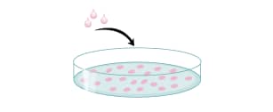
Transfer the cell suspension to a 15 mL centrifuge tube containing >5 mL of pre-warmed hES media.
Centrifuge at 200 x g for 4 minutes.

Remove the supernatant.
Resuspend the pellet in pre-warmed hES media.

Perform a cell count.

Plate the desired number of cells (approximately 0.5-1.0 x 10⁶ cells/60 mm plate) on the iMEF monolayer in hES media supplemented with 4 ng/mL FGF basic.

Replace the media daily.

BG01V cells are licensed from ViaCyte, Inc.
Citations for Irradiated Mouse Embryonic Fibroblasts (5 vials)
R&D Systems personnel manually curate a database that contains references using R&D Systems products. The data collected includes not only links to publications in PubMed, but also provides information about sample types, species, and experimental conditions.
7
Citations: Showing 1 - 7
Filter your results:
Filter by:
-
Transcriptional metabolic reprogramming implements meiotic fate decision in mouse testicular germ cells
Authors: Zhang, X;Liu, Y;Sosa, F;Gunewardena, S;Crawford, PA;Zielen, AC;Orwig, KE;Wang, N;
Cell reports 2023-07-04
-
Early chromosome condensation by XIST builds A-repeat RNA density that facilitates gene silencing
Authors: Valledor, M;Byron, M;Dumas, B;Carone, DM;Hall, LL;Lawrence, JB;
Cell reports 2023-06-28
-
Early chromosome condensation by XIST builds A-repeat RNA density that facilitates gene silencing
Authors: Valledor, M;Byron, M;Dumas, B;Carone, DM;Hall, LL;Lawrence, JB;
Cell reports 2023-06-28
-
Transcriptional profiling of mESC-derived tendon and fibrocartilage cell fate switch
Authors: DA Kaji, AM Montero, R Patel, AH Huang
Nature Communications, 2021-07-09;12(1):4208. 2021-07-09
-
In vitro generation of mouse polarized embryo-like structures from embryonic and trophoblast stem cells
Authors: SE Harrison, B Sozen, M Zernicka-G
Nat Protoc, 2018-07-01;0(0):. 2018-07-01
-
A comparative study of protocols for mouse embryonic stem cell culturing.
Authors: Tamm C, Pijuan Galito S, Anneren C
PLoS ONE, 2013-12-10;8(12):e81156. 2013-12-10
-
Translating dosage compensation to trisomy 21.
Authors: Jiang J, Jing Y, Cost G, Chiang J, Kolpa H, Cotton A, Carone D, Carone B, Shivak D, Guschin D, Pearl J, Rebar E, Byron M, Gregory P, Brown C, Urnov F, Hall L, Lawrence J
Nature, 2013-07-17;500(7462):296-300. 2013-07-17
FAQs
-
If all the cells included in a vial of Irradiated Mouse Embryomic Fibroblasts (Catalog # PSC001) are not used in a single experiment, can the cells be re-frozen and used again?
The Irradiated Mouse Embryonic Fibroblasts will most likely not survive a second freeze-thaw. Freezing the cells after one use is not recommended.
-
How long can Irradiated Mouse Embryonic Fibroblasts (Catalog # PSC001) be cultured before being used in an experiment?
The cells can be cultured for approximately one week. After this period, loss of cell viability is observed.
-
If the vial of Irradiated Mouse Embryonic Fibroblasts (catalog # PSC001) I received is yellow in color, should I still use it?
A yellow vial of catalog # PSC001 may indicate compromised cell viability in the vial. Please contact Technical Services for further assistance.
Reviews for Irradiated Mouse Embryonic Fibroblasts (5 vials)
Average Rating: 4.7 (Based on 7 Reviews)
Have you used Irradiated Mouse Embryonic Fibroblasts (5 vials)?
Submit a review and receive an Amazon gift card.
$25/€18/£15/$25CAN/¥75 Yuan/¥2500 Yen for a review with an image
$10/€7/£6/$10 CAD/¥70 Yuan/¥1110 Yen for a review without an image
Filter by:
I used these cells culture with hiPSC line that were maintained either on an inactivated mouse embryonic fibroblast (MEF) feeder layer
iPSC colonies were identified and separately harvested. Following successive passages onto irradiated MEFs.
I used the mef cells as a feeder cell for stem cell culture. It works good.
Able to culture various hPSC cell lines with success.
