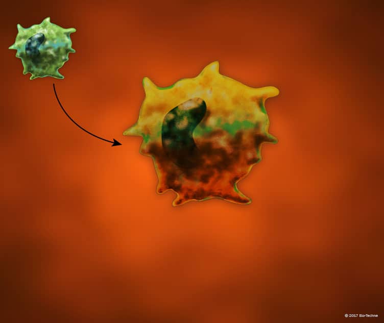
M2b Macrophage Activation State Markers
Click on one of the macrophage activation states shown in the buttons below to see the markers that are commonly used to identify each activated state or view a list of the common macrophage markers used for distinguishing macrophages from other immune cell types.

IL-1 R/TLR Ligands
IL-1 R/TLR Ligands
Overview
M2b Macrophage Activation State Markers Overview
Macrophages are activated following exposure to microbial agonists, cytokines, and growth factors and can display a range of phenotypes depending on the combination of stimuli in their environments. Historically, macrophage activation was suggested to result in polarization toward either a classically activated, M1 pro-inflammatory macrophage phenotype or an alternatively activated M2 anti-inflammatory macrophage phenotype. M2 macrophages have subsequently been divided into different subtypes known as M2a, M2b, M2c, and M2d based on differences in their phenotypes following activation with different stimuli. M2b macrophages are induced following stimulation with immune complexes and IL-1 R or Toll-like Receptor (TLR) agonists, and are associated with increased phagocytic and immunomodulatory activity. M2b macrophages display increased MHC class II and B7-2/CD86 expression, and secrete high amounts of the anti-inflammatory IL-10 cytokine, along with the pro-inflammatory cytokines, IL-1 beta, IL-6, and TNF-alpha.

