M2c Macrophage Activation State Markers
Click on one of the macrophage activation states shown in the buttons below to see the markers that are commonly used to identify each activated state or view a list of the common macrophage markers used for distinguishing macrophages from other immune cell types.

Recommended Products
Overview
M2c Macrophage Activation State Markers Overview
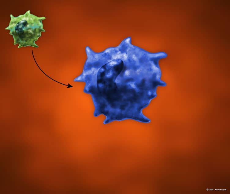
Macrophages are activated following exposure to different stimuli in their environments, which can alter their phenotypes and functions. The classic view of macrophage activation suggests that macrophages undergo either classical or alternative activation in response to different stimuli, leading to the generation of either pro-inflammatory M1 or anti-inflammatory M2 macrophages, respectively. Based on observed differences in the M2 macrophage population following activation with different stimuli, this group of macrophages has subsequently been further divided into the M2a, M2b, M2c, and M2d subtypes. M2c macrophages, also known as deactivated macrophages, are induced following stimulation with IL-10, TGF-beta, and glucocorticoids. M2c macrophages express the cell surface markers, CD163, MMR/CD206, and TLR1, and secrete IL-10 and TGF-beta. Additionally, Arginase 1 (ARG1) is a common mouse M2c macrophage marker.


