The following immunocytochemistry (ICC) protocol has been developed and optimized by R&D Systems’ IHC/ICC laboratory for ICC experiments using cells grown on gelatin-coated glass coverslips.
This ICC protocol provides a basic guide for the preparation, fixation, and fluorescent staining of cells on glass coverslips. Each investigator must determine the precise experimental conditions required to generate a strong and specific signal for each antigen of interest. If R&D Systems’ primary antibodies are employed, please refer to the product data sheets to obtain the recommended working dilutions. In the staining protocol, signal visualization is achieved using R&D Systems’ fluorescent secondary antibodies and reagents. For all other reagents, please follow the manufacturer’s instructions.
Please read ICC protocol in its entirety before starting.
Immunocytochemistry Protocol for Coverslip Preparation Using Gelatin
Many cultured cell types do not adhere well to glass coverslips. Adding a thin layer of gelatin to a coverslip enhances the adhesion of cultured cells to glass.
Reagents Required
- Gelatin-coating solution: 0.1% gelatin in deionized H2O
Materials
- Coverslips (sterilized)
- Cell culture plate (6- or 24-well)
Immunocytochemistry Coverslip Protocol
- Place sterilized coverslips into the wells of a 24-well plate.
- Add 400 µL of the gelatin-coating solution, and incubate the coverslips for 10 minutes at room temperature.
- Remove the gelatin-coating solution, and air dry the coverslips for 15 minutes.
- The dried coverslips can now be stored at room temperature until use.
Immunocytochemistry Protocol for the Preparation & Fixation of Cells on Coverslips
Reagents Required
- 1X PBS: 0.145 M NaCl, 0.0027 M KCl, 0.0081 M Na2HPO4, 0.0015 M KH2PO4, pH 7.4
- Formaldehyde Fixative Solution: 2-4% paraformaldehyde in PBS
- Wash buffer: 0.1% BSA in 1X PBS
- Cell culture medium
Materials
- Gelatin-coated coverslips in a 24-well plate
Immunocytochemistry Preparation & Fixation Protocol
- Culture cells by adding 500 µL of culture media containing approximately 5000 cells to the wells of a cell culture plate containing gelatin-coated coverslips.
- When cells have reached the desired density/age, remove the culture media from each well and wash twice with PBS.
- Add 300-400 µL of 2-4% Formaldehyde Fixative Solution to each well, and incubate for 20 minutes at room temperature.
Note: Some cell types can be damaged by the change in surface tension that occurs when the culture medium is entirely removed and replaced with wash buffer. If this is the case, pre-fix the cells by adding 500 µL of 4% Formaldehyde Fixative Solution directly into the culture medium. After 2 minutes, replace the pre-fixation culture medium with 300-400 µL of 2% Formaldehyde Fixative Solution and incubate, for 20 minutes at room temperature. - Wash the wells twice with PBS and cover with 400 µL of wash buffer. The coverslips can be stored at 2-8 °C for up to 3 months or they may be stained immediately.
Note: Fixation can result in hydrophobic cross-linking of tissue proteins. The time, temperature, pH, and fixative used will determine the degree of cross-linking. Once the fixation protocol has been optimized, the same procedure should be used consistently.
Immunocytochemistry Protocol for Fluorescent Staining of Cultured Cells on Coverslips
Reagents Required
- Primary Antibodies
- Blocking buffer: 10% normal donkey serum, 0.3% Triton® X-100
- DAPI (4',6-diamidino-2-phenylindole) solution: Add 1 µL of 14.3 mM stock for every 5 mL of PBS. Store any unused DAPI at 2-8 °C, wrapped in aluminum foil
- Deionized H2O
- Dilution buffer: 1X PBS, 1% bovine serum albumin (BSA), 1% normal donkey serum, 0.3% Triton X-100, and 0.01% sodium azide
- Anti-fade mounting medium
- Conjugated secondary reagents (or equivalent)
- 1X PBS: 0.145 M NaCl, 0.0027 M KCl, 0.0081 M Na2HPO4, 0.0015 M KH2PO4, pH 7.4
- Wash buffer: 0.1% BSA in 1X PBS
Materials
- Cell-covered coverslips in a 6- or 24-well plate
- Fine tweezers
Immunocytochemsitry Protocol
Note: This protocol is optimized for cells grown on coverslips in a 6- or 24-well plate but can be adapted accordingly.
- Wash the coverslips containing the fixed cells two times in 400 µL of wash buffer.
- Block non-specific staining by adding 400 µL of blocking buffer and incubate for 45 minutes at room temperature.
- Remove blocking buffer. No rinsing is necessary.
- Dilute the unconjugated primary antibody (or fluorescence-conjugated primary) in dilution buffer according to the manufacturer’s instructions. For fluorescent ICC staining of cells on coverslips using R&D Systems antibodies, it is recommended to incubate at room temperature for 1 hour. Alternatively, incubate overnight at 2-8 °C.
Note: Appropriate controls are critical for the accurate interpretation of IHC/ICC results. All IHC/ICC experiments should include a negative control using the incubation buffer with no primary antibody to identify non-specific staining of the secondary reagents. Additional controls can be employed to support the specificity of staining generated by the primary antibody. These include absorption controls, isotype controls (for monoclonal primary antibodies), and tissue type controls. - Wash two times in 400 µL of wash buffer. If using a primary antibody with a direct fluorescent conjugate, go to step 8.
- Dilute the secondary antibody in dilution buffer according to the manufacturer’s instructions. Add 400 µL to the wells, and incubate at room temperature for 1 hour in the dark. From this step forward samples should be protected from light.
Note: NorthernLightsTMfluorescent secondary antibodies and streptavidin conjugates are bright, resistant to photobleaching, and are ideal for multi-color fluorescence microscopy.
Note: If a biotinylated antibody was used in step 4, apply Streptavidin-conjugated to fluorescent probe in step 6. - Rinse two times in 400 µL of wash buffer.
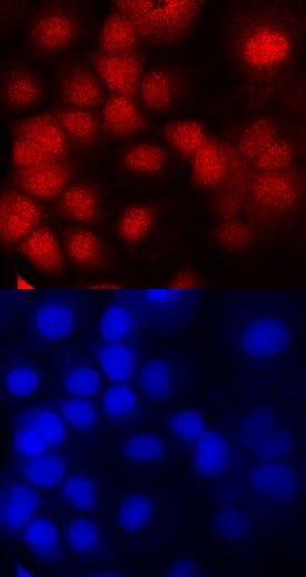
- Add 300 µL of the diluted DAPI solution to each well, and incubate 2-5 minutes at room temperature. DAPI binds to DNA and is a convenient nuclear counterstain. It has an absorption maximum at 358 nm and fluoresces blue at an emission maximum of 461 nm.
Note: DAPI counterstain can obscure visualization of targets localized in cell nuclei. - Rinse once with PBS and once with water.
- Carefully remove the coverslips from the wells and blot to remove any excess water. Dispense 1 drop of anti-fade mounting medium onto the microscope slide per coverslip. Mount the coverslip with the cells facing towards the microscope slide.
- Visualize using a fluorescence microscope and filter sets appropriate for the label used. Slides can also be stored in a slide box at < -20 °C for later examination.
Note: Initial IHC/ICC studies often require further optimization and/or additional troubleshooting steps.
Triton is a registered trademark of Dow Chemical.
Related IHC/ICC Resources
- Troubleshooting Guide for Immunohistochemistry and Immunocytochemsitry
- Protocol for Making a 4% Formaldehyde Solution in PBS
- IHC/ICC Protocol Guide
- Designing a Successful IHC/ICC Experiment
- All IHC/ICC protocols


